 Movie
Movie Controller


+ Open data

- Basic information
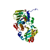
| Entry | Database: PDB / ID: 5t8u | |||||||||
|---|---|---|---|---|---|---|---|---|---|---|
| Title | Crystal structure of P. falciparum LipL1 in complex lipoate | |||||||||
 Components Components | Lipoate-protein ligase 1 Lipoate–protein ligase Lipoate–protein ligase | |||||||||
 Keywords Keywords | LIGASE / Lipoylation | |||||||||
| Function / homology |  Function and homology information Function and homology informationGlyoxylate metabolism and glycine degradation / lipoyltransferase activity / lipoate-protein ligase / lipoate-protein ligase activity / protein lipoylation / lipid metabolic process / mitochondrion / ATP binding / cytoplasmSimilarity search - Function | |||||||||
| Biological species |  Plasmodium falciparum (malaria parasite P. falciparum) Plasmodium falciparum (malaria parasite P. falciparum) | |||||||||
| Method | X-RAY DIFFRACTION / SYNCHROTRON / MOLECULAR REPLACEMENT / Resolution: 2.324 Å | |||||||||
 Authors Authors | Guerra, A.J. / Afanador, G.A. / Prigge, S.T. | |||||||||
| Funding support |  United States, 2items United States, 2items
| |||||||||
 Citation Citation | Journal: Proteins / Year: 2017 Title: Crystal structure of lipoate-bound lipoate ligase 1, LipL1, from Plasmodium falciparum. Authors: Guerra, A.J. / Afanador, G.A. / Prigge, S.T. | |||||||||
| History |
|
- Structure visualization
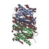
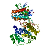
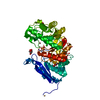
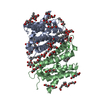
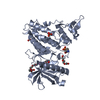
| Structure viewer | Molecule: MolmilJmol/JSmol |
|---|
- Downloads & links
-Download
| PDBx/mmCIF format | 5t8u.cif.gz | 400.5 KB | Display | PDBx/mmCIF format |
|---|---|---|---|---|
| PDB format | pdb5t8u.ent.gz | 339.6 KB | Display | PDB format |
| PDBx/mmJSON format | 5t8u.json.gz | Tree view | PDBx/mmJSON format | |
| Others |  Other downloads Other downloads |
-Validation report
| Arichive directory | https://data.pdbj.org/pub/pdb/validation_reports/t8/5t8uftp://data.pdbj.org/pub/pdb/validation_reports/t8/5t8u | HTTPS FTP |
|---|
-Related structure data
| Related structure data |  3a7aS S: Starting model for refinement |
|---|---|
| Similar structure data |


-Links
 PDBj
PDBj
- Assembly
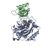
| Deposited unit | 
| ||||||||
|---|---|---|---|---|---|---|---|---|---|
| 1 | 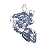
| ||||||||
| 2 | 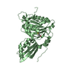
| ||||||||
| Unit cell |
|
-Components
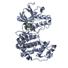
| #1: Protein | Lipoate–protein ligase Mass: 42204.832 Da / Num. of mol.: 2 Source method: isolated from a genetically manipulated source Source: (gene. exp.) Plasmodium falciparum (malaria parasite P. falciparum)Strain: isolate 3D7 / Gene: PF3D7_1314600 / Plasmid: pMALcHT / Production host:  Escherichia coli (E. coli) / Strain (production host): BL21 STAR (DE3) / References: UniProt: Q8IEG9, EC: 2.7.7.63 Escherichia coli (E. coli) / Strain (production host): BL21 STAR (DE3) / References: UniProt: Q8IEG9, EC: 2.7.7.63#2: Chemical | Lipoic acid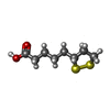  Mass: 206.326 Da / Num. of mol.: 2 / Source method: obtained synthetically / Formula: C8H14O2S2 Mass: 206.326 Da / Num. of mol.: 2 / Source method: obtained synthetically / Formula: C8H14O2S2#3: Water | ChemComp-HOH / | Water Mass: 18.015 Da / Num. of mol.: 84 / Source method: isolated from a natural source / Formula: H2O Mass: 18.015 Da / Num. of mol.: 84 / Source method: isolated from a natural source / Formula: H2O |
|---|
-Experimental details
-Experiment
| Experiment | Method: X-RAY DIFFRACTION / Number of used crystals: 1 |
|---|
- Sample preparation
| Crystal | Density Matthews: 3.26 Å3/Da / Density % sol: 62.23 % |
|---|---|
| Crystal grow | Temperature: 293 K / Method: vapor diffusion, sitting drop / pH: 7 / Details: 1.5M Ammonium Sulfate, 20% Ethylene Glycol |
-Data collection
| Diffraction | Mean temperature: 100 K |
|---|---|
| Diffraction source | Source: SYNCHROTRON / Site: SSRL / Beamline: BL7-1 / Wavelength: 1.127085 Å |
| Detector | Type: ADSC QUANTUM 315r / Detector: CCD / Date: May 25, 2015 |
| Radiation | Protocol: SINGLE WAVELENGTH / Monochromatic (M) / Laue (L): M / Scattering type: x-ray |
| Radiation wavelength | Wavelength: 1.127085 Å / Relative weight: 1 |
| Reflection | Resolution: 2.324→48.574 Å / Num. obs: 48667 / % possible obs: 96 % / Redundancy: 5.9 % / Biso Wilson estimate: 53.09 Å2 / CC1/2: 0.998 / Rmerge(I) obs: 0.1391 / Net I/σ(I): 9.9 |
- Processing
| Software |
| |||||||||||||||||||||||||||||||||||||||||||||||||||||||||||||||||||||||||||||||||||||||||||||||||||||||||||||||||||||||||||||
|---|---|---|---|---|---|---|---|---|---|---|---|---|---|---|---|---|---|---|---|---|---|---|---|---|---|---|---|---|---|---|---|---|---|---|---|---|---|---|---|---|---|---|---|---|---|---|---|---|---|---|---|---|---|---|---|---|---|---|---|---|---|---|---|---|---|---|---|---|---|---|---|---|---|---|---|---|---|---|---|---|---|---|---|---|---|---|---|---|---|---|---|---|---|---|---|---|---|---|---|---|---|---|---|---|---|---|---|---|---|---|---|---|---|---|---|---|---|---|---|---|---|---|---|---|---|---|
| Refinement | Method to determine structure: MOLECULAR REPLACEMENT Starting model: 3A7A Resolution: 2.324→48.574 Å / SU ML: 0.55 / Cross valid method: FREE R-VALUE / σ(F): 1.33 / Phase error: 42.38 / Stereochemistry target values: ML
| |||||||||||||||||||||||||||||||||||||||||||||||||||||||||||||||||||||||||||||||||||||||||||||||||||||||||||||||||||||||||||||
| Solvent computation | Shrinkage radii: 0.9 Å / VDW probe radii: 1.11 Å / Solvent model: FLAT BULK SOLVENT MODEL | |||||||||||||||||||||||||||||||||||||||||||||||||||||||||||||||||||||||||||||||||||||||||||||||||||||||||||||||||||||||||||||
| Refinement step | Cycle: LAST / Resolution: 2.324→48.574 Å
| |||||||||||||||||||||||||||||||||||||||||||||||||||||||||||||||||||||||||||||||||||||||||||||||||||||||||||||||||||||||||||||
| Refine LS restraints |
| |||||||||||||||||||||||||||||||||||||||||||||||||||||||||||||||||||||||||||||||||||||||||||||||||||||||||||||||||||||||||||||
| LS refinement shell |
| |||||||||||||||||||||||||||||||||||||||||||||||||||||||||||||||||||||||||||||||||||||||||||||||||||||||||||||||||||||||||||||
| Refinement TLS params. | Method: refined / Refine-ID: X-RAY DIFFRACTION
| |||||||||||||||||||||||||||||||||||||||||||||||||||||||||||||||||||||||||||||||||||||||||||||||||||||||||||||||||||||||||||||
| Refinement TLS group |
|
