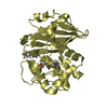
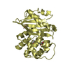
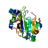
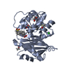
Mass: 11733.242 Da / Num. of mol.: 1 / Fragment: catalytic domain Source method: isolated from a genetically manipulated source Details: The cloned fragment starts with V3033 of the original sequence. The purified complex was trypsin-treated and the sequence of most likely cleavage product is provided. Source: (gene. exp.) Escherichia coli (E. coli) / Gene: eco1013 / Plasmid: pMCSG81 / Production host: Escherichia coli (E. coli) / Strain (production host): BL21(DE3) / References: UniProt: Q1RPM1
#2: Protein
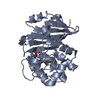
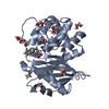
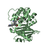
CdiIimmunityprotein
Mass: 15051.525 Da / Num. of mol.: 1 Source method: isolated from a genetically manipulated source Source: (gene. exp.) Escherichia coli (E. coli) / Gene: AGA26_04870 / Plasmid: pMCSG81 / Production host: Escherichia coli (E. coli) / Strain (production host): BL21(DE3) / References: UniProt: A0A0B0W5A7
Monochromator: SI (111) / Protocol: SINGLE WAVELENGTH / Monochromatic (M) / Laue (L): M / Scattering type: x-ray
Radiation wavelength
Wavelength: 0.9793 Å / Relative weight: 1
Reflection
Resolution: 2→30 Å / Num. obs: 17148 / % possible obs: 99.8 % / Observed criterion σ(I): -3 / Redundancy: 6 % / Rmerge(I) obs: 0.104 / Net I/σ(I): 16.17
Reflection shell
Resolution: 2→2.03 Å / Redundancy: 5.1 % / Rmerge(I) obs: 0.743 / Mean I/σ(I) obs: 1.94 / % possible all: 99.5
-
Processing
Software
Name
Version
Classification
REFMAC
5.8.0155
refinement
HKL-3000
datareduction
HKL-3000
datascaling
HKL-3000
phasing
SBC-Collect
datacollection
Refinement
Method to determine structure: SAD / Resolution: 2→30 Å / Cor.coef. Fo:Fc: 0.956 / Cor.coef. Fo:Fc free: 0.917 / SU B: 8.826 / SU ML: 0.119 / Cross valid method: THROUGHOUT / ESU R: 0.183 / ESU R Free: 0.168 / Stereochemistry target values: MAXIMUM LIKELIHOOD / Details: HYDROGENS HAVE BEEN ADDED IN THE RIDING POSITIONS
Rfactor
Num. reflection
% reflection
Selection details
Rfree
0.23396
799
4.8 %
RANDOM
Rwork
0.18278
-
-
-
obs
0.18507
15909
97.43 %
-
Solvent computation
Ion probe radii: 0.8 Å / Shrinkage radii: 0.8 Å / VDW probe radii: 1.2 Å / Solvent model: MASK
 Movie
Movie Controller


 Open data
 Basic information
 Components
 Keywords
 TOXIN /
  Function and homology information
 Authors
 United States, 1items
  Citation
 Structure visualization
 Downloads & links
 Other downloads

 PDBj
PDBj Assembly
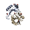

 Mass: 59.044 Da / Num. of mol.: 5 / Source method: obtained synthetically / Formula: C2H3O2
 Mass: 18.015 Da / Num. of mol.: 106 / Source method: isolated from a natural source / Formula: H2O
 Sample preparation
 Processing