| Entry | Database: PDB / ID: 5swm
|
|---|
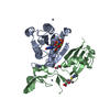
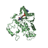
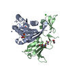
| Title | BACILLUS HALODURANS RNASE H MUTANT D132N IN COMPLEX WITH 12-MER FRNA/DNA HYBRID |
|---|
 Components Components | - DNA (12-MER)
- RNA (12-MER)
- Ribonuclease H

|
|---|
 Keywords Keywords | HYDROLASE/RNA/DNA / RNASE H / RNA/DNA HYBRID / HYDROLASE-RNA-DNA COMPLEX |
|---|
| Function / homology |  Function and homology information Function and homology information |
|---|
| Biological species |  Bacillus halodurans (bacteria) Bacillus halodurans (bacteria)
synthetic construct (others) |
|---|
| Method | X-RAY DIFFRACTION / SYNCHROTRON / MOLECULAR REPLACEMENT / Resolution: 1.5 Å |
|---|
 Authors Authors | Pallan, P.S. / Egli, M. |
|---|
| Funding support |  United States, 1items United States, 1items | Organization | Grant number | Country |
|---|
| National Institutes of Health/National Institute of General Medical Sciences (NIH/NIGMS) | R01 GM055237 | United States |
|
|---|
 Citation Citation | Journal: Biochemistry / Year: 2016
Title: Limits of RNA 2'-OH Mimicry by Fluorine: Crystal Structure of Bacillus halodurans RNase H Bound to a 2'-FRNA:DNA Hybrid.
Authors: Pallan, P.S. / Prakash, T.P. / de Leon, A.R. / Egli, M. |
|---|
| History | | Deposition | Aug 8, 2016 | Deposition site: RCSB / Processing site: RCSB |
|---|
| Revision 1.0 | Sep 21, 2016 | Provider: repository / Type: Initial release |
|---|
| Revision 1.1 | Oct 12, 2016 | Group: Database references |
|---|
| Revision 1.2 | Sep 27, 2017 | Group: Author supporting evidence / Database references / Derived calculations
Category: citation / pdbx_audit_support / pdbx_struct_oper_list
Item: _citation.journal_id_CSD / _pdbx_audit_support.funding_organization / _pdbx_struct_oper_list.symmetry_operation |
|---|
| Revision 1.3 | Dec 25, 2019 | Group: Author supporting evidence / Category: pdbx_audit_support / Item: _pdbx_audit_support.funding_organization |
|---|
| Revision 1.4 | Oct 4, 2023 | Group: Data collection / Database references ...Data collection / Database references / Derived calculations / Refinement description
Category: chem_comp_atom / chem_comp_bond ...chem_comp_atom / chem_comp_bond / database_2 / pdbx_initial_refinement_model / struct_conn / struct_conn_type
Item: _database_2.pdbx_DOI / _database_2.pdbx_database_accession ..._database_2.pdbx_DOI / _database_2.pdbx_database_accession / _struct_conn.conn_type_id / _struct_conn.id / _struct_conn.pdbx_dist_value / _struct_conn.pdbx_leaving_atom_flag / _struct_conn.pdbx_ptnr1_label_alt_id / _struct_conn.pdbx_ptnr2_label_alt_id / _struct_conn.ptnr1_auth_asym_id / _struct_conn.ptnr1_auth_comp_id / _struct_conn.ptnr1_auth_seq_id / _struct_conn.ptnr1_label_asym_id / _struct_conn.ptnr1_label_atom_id / _struct_conn.ptnr1_label_comp_id / _struct_conn.ptnr1_label_seq_id / _struct_conn.ptnr2_auth_asym_id / _struct_conn.ptnr2_auth_comp_id / _struct_conn.ptnr2_auth_seq_id / _struct_conn.ptnr2_label_asym_id / _struct_conn.ptnr2_label_atom_id / _struct_conn.ptnr2_label_comp_id / _struct_conn.ptnr2_label_seq_id / _struct_conn_type.id |
|---|
|
|---|
 Movie
Movie Controller

 Yorodumi
 Open data
 Basic information
 Structure visualization
 Downloads & links
 Other downloads


 PDBj
PDBj


 Assembly


 Mass: 22.990 Da / Num. of mol.: 1 / Source method: isolated from a natural source / Formula: Na
 Mass: 35.453 Da / Num. of mol.: 1 / Source method: isolated from a natural source / Formula: Cl
 Mass: 106.120 Da / Num. of mol.: 1 / Source method: isolated from a natural source / Formula: C4H10O3
 Mass: 92.094 Da / Num. of mol.: 1 / Source method: isolated from a natural source / Formula: C3H8O3
 Sample preparation
 Processing