 Movie
Movie Controller

[English] 日本語
 Yorodumi
- PDB-5qr2: PanDDA analysis group deposition -- Crystal Structure of human AL... -
+ Open data

- Basic information
| Entry | Database: PDB / ID: 5qr2 | ||||||
|---|---|---|---|---|---|---|---|
| Title | PanDDA analysis group deposition -- Crystal Structure of human ALAS2A in complex with Z1348371854 | ||||||
 Components Components | 5-aminolevulinate synthase, erythroid-specific, mitochondrial Aminolevulinic acid synthase Aminolevulinic acid synthase | ||||||
 Keywords Keywords | TRANSFERASE / SGC - Diamond I04-1 fragment screening / PanDDA / XChemExplorer | ||||||
| Function / homology |  Function and homology information5-aminolevulinate synthase / 5-aminolevulinate synthase activity / hemoglobin biosynthetic process / intracellular oxygen homeostasis / protoporphyrinogen IX biosynthetic process / Heme biosynthesis / erythrocyte development / heme biosynthetic process / erythrocyte differentiation / pyridoxal phosphate binding ...5-aminolevulinate synthase / 5-aminolevulinate synthase activity / hemoglobin biosynthetic process / intracellular oxygen homeostasis / protoporphyrinogen IX biosynthetic process / Heme biosynthesis / erythrocyte development / heme biosynthetic process / erythrocyte differentiation / pyridoxal phosphate binding / mitochondrial inner membrane / intracellular iron ion homeostasis / response to hypoxia / mitochondrial matrix / mitochondrion Function and homology information5-aminolevulinate synthase / 5-aminolevulinate synthase activity / hemoglobin biosynthetic process / intracellular oxygen homeostasis / protoporphyrinogen IX biosynthetic process / Heme biosynthesis / erythrocyte development / heme biosynthetic process / erythrocyte differentiation / pyridoxal phosphate binding ...5-aminolevulinate synthase / 5-aminolevulinate synthase activity / hemoglobin biosynthetic process / intracellular oxygen homeostasis / protoporphyrinogen IX biosynthetic process / Heme biosynthesis / erythrocyte development / heme biosynthetic process / erythrocyte differentiation / pyridoxal phosphate binding / mitochondrial inner membrane / intracellular iron ion homeostasis / response to hypoxia / mitochondrial matrix / mitochondrionSimilarity search - Function | ||||||
| Biological species |  Homo sapiens (human) Homo sapiens (human) | ||||||
| Method | X-RAY DIFFRACTION / SYNCHROTRON / FOURIER SYNTHESIS / molecular replacement / Resolution: 1.66 Å | ||||||
 Authors Authors | Bezerra, G.A. / Foster, W. / Bailey, H. / Shrestha, L. / Krojer, T. / Talon, R. / Brandao-Neto, J. / Douangamath, A. / Nicola, B.B. / von Delft, F. ...Bezerra, G.A. / Foster, W. / Bailey, H. / Shrestha, L. / Krojer, T. / Talon, R. / Brandao-Neto, J. / Douangamath, A. / Nicola, B.B. / von Delft, F. / Arrowsmith, C.H. / Edwards, A. / Bountra, C. / Brennan, P.E. / Yue, W.W. | ||||||
 Citation Citation | Journal: To Be Published Title: PanDDA analysis group deposition Authors: Bezerra, G.A. / Foster, W. / Bailey, H. / Shrestha, L. / Krojer, T. / Talon, R. / Brandao-Neto, J. / Douangamath, A. / Nicola, B.B. / von Delft, F. / Arrowsmith, C.H. / Edwards, A. / ...Authors: Bezerra, G.A. / Foster, W. / Bailey, H. / Shrestha, L. / Krojer, T. / Talon, R. / Brandao-Neto, J. / Douangamath, A. / Nicola, B.B. / von Delft, F. / Arrowsmith, C.H. / Edwards, A. / Bountra, C. / Brennan, P.E. / Yue, W.W. | ||||||
| History |
|
- Structure visualization
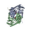
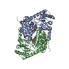
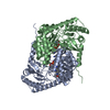
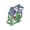
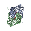
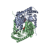
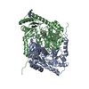
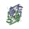
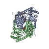
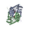
| Structure viewer | Molecule: MolmilJmol/JSmol |
|---|
- Downloads & links
-Download
| PDBx/mmCIF format | 5qr2.cif.gz | 182.7 KB | Display | PDBx/mmCIF format |
|---|---|---|---|---|
| PDB format | pdb5qr2.ent.gz | 143.6 KB | Display | PDB format |
| PDBx/mmJSON format | 5qr2.json.gz | Tree view | PDBx/mmJSON format | |
| Others |  Other downloads Other downloads |
-Validation report
| Arichive directory | https://data.pdbj.org/pub/pdb/validation_reports/qr/5qr2ftp://data.pdbj.org/pub/pdb/validation_reports/qr/5qr2 | HTTPS FTP |
|---|
-Group deposition
| ID | G_1002078 (25 entries) |
|---|---|
| Title | PanDDA analysis group deposition |
| Type | changed state |
| Description | human 5'-Aminolevulinate synthase erythroid-specific (ALAS2) screened against the DSi Poised Fragment Library by X-ray Crystallography at the XChem facility of Diamond Light Source beamline I04-1 |


-Related structure data
| Related structure data | 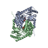 6hrhS S: Starting model for refinement |
|---|---|
| Similar structure data |


-Links
 PDBj
PDBj

- Assembly
| Deposited unit | 
| ||||||||
|---|---|---|---|---|---|---|---|---|---|
| 1 |
| ||||||||
| Unit cell |
| ||||||||
| Components on special symmetry positions |
|

-Components
| #1: Protein | Aminolevulinic acid synthase / ALAS-E / 5-aminolevulinic acid synthase 2 / Delta-ALA synthase 2 / Delta-aminolevulinate synthase 2 Mass: 52250.555 Da / Num. of mol.: 2 Source method: isolated from a genetically manipulated source Source: (gene. exp.) Homo sapiens (human) / Gene: ALAS2, ALASE, ASB / Production host:   Spodoptera frugiperda (fall armyworm) / References: UniProt: P22557, 5-aminolevulinate synthase Spodoptera frugiperda (fall armyworm) / References: UniProt: P22557, 5-aminolevulinate synthase#2: Chemical | Pyridoxal phosphate  Mass: 247.142 Da / Num. of mol.: 2 / Source method: obtained synthetically / Formula: C8H10NO6P Mass: 247.142 Da / Num. of mol.: 2 / Source method: obtained synthetically / Formula: C8H10NO6P#3: Chemical | ChemComp-NTG / |   Mass: 203.240 Da / Num. of mol.: 1 / Source method: obtained synthetically / Formula: C11H13N3O / Feature type: SUBJECT OF INVESTIGATION Mass: 203.240 Da / Num. of mol.: 1 / Source method: obtained synthetically / Formula: C11H13N3O / Feature type: SUBJECT OF INVESTIGATION#4: Water | ChemComp-HOH / | Water Mass: 18.015 Da / Num. of mol.: 248 / Source method: isolated from a natural source / Formula: H2O Mass: 18.015 Da / Num. of mol.: 248 / Source method: isolated from a natural source / Formula: H2OHas ligand of interest | Y | |
|---|
-Experimental details
-Experiment
| Experiment | Method: X-RAY DIFFRACTION / Number of used crystals: 1 |
|---|
- Sample preparation
| Crystal | Density Matthews: 2.32 Å3/Da / Density % sol: 46.92 % |
|---|---|
| Crystal grow | Temperature: 298 K / Method: vapor diffusion, sitting drop / pH: 7.5 Details: 0.1 M HEPES pH 7.5, 0.2 M magnesium chloride, 20% PEG3350 |
-Data collection
| Diffraction | Mean temperature: 100 K | ||||||||||||||||||||||||
|---|---|---|---|---|---|---|---|---|---|---|---|---|---|---|---|---|---|---|---|---|---|---|---|---|---|
| Diffraction source | Source: SYNCHROTRON / Site: Diamond  / Beamline: I04-1 / Wavelength: 0.91587 Å / Beamline: I04-1 / Wavelength: 0.91587 Å | ||||||||||||||||||||||||
| Detector | Type: DECTRIS PILATUS 6M / Detector: PIXEL / Date: Apr 9, 2019 | ||||||||||||||||||||||||
| Radiation | Protocol: SINGLE WAVELENGTH / Scattering type: x-ray | ||||||||||||||||||||||||
| Radiation wavelength | Wavelength: 0.91587 Å / Relative weight: 1 | ||||||||||||||||||||||||
| Reflection | Resolution: 1.662→79.91 Å / Num. obs: 70623 / % possible obs: 91.1 % / Redundancy: 3.4 % / CC1/2: 0.998 / Rpim(I) all: 0.05 / Rrim(I) all: 0.093 / Net I/σ(I): 9.6 / Num. measured all: 242960 | ||||||||||||||||||||||||
| Reflection shell | Diffraction-ID: 1 / Redundancy: 3.4 %
|
-Phasing
| Phasing | Method: molecular replacement |
|---|
- Processing
| Software |
| |||||||||||||||||||||||||||||||||||||||||||||||||||||||||||||||||||||||||||
|---|---|---|---|---|---|---|---|---|---|---|---|---|---|---|---|---|---|---|---|---|---|---|---|---|---|---|---|---|---|---|---|---|---|---|---|---|---|---|---|---|---|---|---|---|---|---|---|---|---|---|---|---|---|---|---|---|---|---|---|---|---|---|---|---|---|---|---|---|---|---|---|---|---|---|---|---|
| Refinement | Method to determine structure: FOURIER SYNTHESIS Starting model: 6hrh Resolution: 1.66→79.91 Å / Cor.coef. Fo:Fc: 0.941 / Cor.coef. Fo:Fc free: 0.929 / SU B: 3.097 / SU ML: 0.1 / Cross valid method: THROUGHOUT / σ(F): 0 / ESU R: 0.175 / ESU R Free: 0.15 / Stereochemistry target values: MAXIMUM LIKELIHOOD Details: HYDROGENS HAVE BEEN ADDED IN THE RIDING POSITIONS U VALUES : REFINED INDIVIDUALLY
| |||||||||||||||||||||||||||||||||||||||||||||||||||||||||||||||||||||||||||
| Solvent computation | Ion probe radii: 0.8 Å / Shrinkage radii: 0.8 Å / VDW probe radii: 1.2 Å / Solvent model: MASK | |||||||||||||||||||||||||||||||||||||||||||||||||||||||||||||||||||||||||||
| Displacement parameters | Biso max: 77.56 Å2 / Biso mean: 20.945 Å2 / Biso min: 5.62 Å2
| |||||||||||||||||||||||||||||||||||||||||||||||||||||||||||||||||||||||||||
| Refinement step | Cycle: final / Resolution: 1.66→79.91 Å
| |||||||||||||||||||||||||||||||||||||||||||||||||||||||||||||||||||||||||||
| Refine LS restraints |
| |||||||||||||||||||||||||||||||||||||||||||||||||||||||||||||||||||||||||||
| LS refinement shell | Resolution: 1.662→1.705 Å / Total num. of bins used: 20
|
