 Movie
Movie Controller


+ Open data

- Basic information
| Entry | Database: PDB / ID: 5qcg | |||||||||
|---|---|---|---|---|---|---|---|---|---|---|
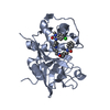
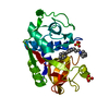
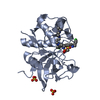
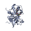
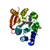
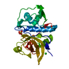
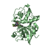
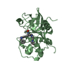
| Title | Crystal structure of human Cathepsin-S with bound ligand | |||||||||
 Components Components | Cathepsin S | |||||||||
 Keywords Keywords | HYDROLASE / D3R / Cathepsin S / Ligand Docking | |||||||||
| Function / homology |  Function and homology informationcathepsin S / basement membrane disassembly / positive regulation of cation channel activity / antigen processing and presentation of peptide antigen / endolysosome lumen / response to acidic pH / cellular response to thyroid hormone stimulus / Trafficking and processing of endosomal TLR / proteoglycan binding / Assembly of collagen fibrils and other multimeric structures ...cathepsin S / basement membrane disassembly / positive regulation of cation channel activity / antigen processing and presentation of peptide antigen / endolysosome lumen / response to acidic pH / cellular response to thyroid hormone stimulus / Trafficking and processing of endosomal TLR / proteoglycan binding / Assembly of collagen fibrils and other multimeric structures / toll-like receptor signaling pathway / cysteine-type endopeptidase activator activity involved in apoptotic process / fibronectin binding / antigen processing and presentation / collagen catabolic process / extracellular matrix disassembly / laminin binding / phagocytic vesicle / collagen binding / MHC class II antigen presentation / Degradation of the extracellular matrix / lysosomal lumen / proteolysis involved in protein catabolic process / positive regulation of apoptotic signaling pathway / Endosomal/Vacuolar pathway / protein processing / antigen processing and presentation of exogenous peptide antigen via MHC class II / late endosome / tertiary granule lumen / collagen-containing extracellular matrix / adaptive immune response / ficolin-1-rich granule lumen / lysosome / immune response / cysteine-type endopeptidase activity / serine-type endopeptidase activity / intracellular membrane-bounded organelle / Neutrophil degranulation / proteolysis / extracellular space / extracellular region Function and homology informationcathepsin S / basement membrane disassembly / positive regulation of cation channel activity / antigen processing and presentation of peptide antigen / endolysosome lumen / response to acidic pH / cellular response to thyroid hormone stimulus / Trafficking and processing of endosomal TLR / proteoglycan binding / Assembly of collagen fibrils and other multimeric structures ...cathepsin S / basement membrane disassembly / positive regulation of cation channel activity / antigen processing and presentation of peptide antigen / endolysosome lumen / response to acidic pH / cellular response to thyroid hormone stimulus / Trafficking and processing of endosomal TLR / proteoglycan binding / Assembly of collagen fibrils and other multimeric structures / toll-like receptor signaling pathway / cysteine-type endopeptidase activator activity involved in apoptotic process / fibronectin binding / antigen processing and presentation / collagen catabolic process / extracellular matrix disassembly / laminin binding / phagocytic vesicle / collagen binding / MHC class II antigen presentation / Degradation of the extracellular matrix / lysosomal lumen / proteolysis involved in protein catabolic process / positive regulation of apoptotic signaling pathway / Endosomal/Vacuolar pathway / protein processing / antigen processing and presentation of exogenous peptide antigen via MHC class II / late endosome / tertiary granule lumen / collagen-containing extracellular matrix / adaptive immune response / ficolin-1-rich granule lumen / lysosome / immune response / cysteine-type endopeptidase activity / serine-type endopeptidase activity / intracellular membrane-bounded organelle / Neutrophil degranulation / proteolysis / extracellular space / extracellular regionSimilarity search - Function | |||||||||
| Biological species |  Homo sapiens (human) Homo sapiens (human) | |||||||||
| Method | X-RAY DIFFRACTION / MOLECULAR REPLACEMENT / molecular replacement / Resolution: 2.7 Å | |||||||||
| Model details | Structures re-refined for D3R docking challenge | |||||||||
 Authors Authors | Bembenek, S.D. / Ameriks, M.K. / Mirzadegan, T. / Yang, H. / Shao, C. / Burley, S.K. | |||||||||
 Citation Citation | Journal: To be published Title: Crystal structure of human Cathepsin-S with bound ligand Authors: Bembenek, S.D. / Ameriks, M.K. / Mirzadegan, T. / Yang, H. / Shao, C. / Burley, S.K. | |||||||||
| History |
|
- Structure visualization
| Structure viewer | Molecule: MolmilJmol/JSmol |
|---|
- Downloads & links
-Download
| PDBx/mmCIF format | 5qcg.cif.gz | 98 KB | Display | PDBx/mmCIF format |
|---|---|---|---|---|
| PDB format | pdb5qcg.ent.gz | 79.4 KB | Display | PDB format |
| PDBx/mmJSON format | 5qcg.json.gz | Tree view | PDBx/mmJSON format | |
| Others |  Other downloads Other downloads |
-Validation report
| Arichive directory | https://data.pdbj.org/pub/pdb/validation_reports/qc/5qcgftp://data.pdbj.org/pub/pdb/validation_reports/qc/5qcg | HTTPS FTP |
|---|
-Group deposition
| ID | G_1002040 (26 entries) |
|---|---|
| Title | Ligand binding to Cathepsin S |
| Type | undefined |
| Description | Ligand binding to Cathepsin S |


-Related structure data
| Similar structure data |
|---|


-Links
 PDBj
PDBj


- Assembly
| Deposited unit | 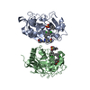
| ||||||||
|---|---|---|---|---|---|---|---|---|---|
| 1 | 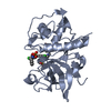
| ||||||||
| 2 | 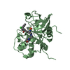
| ||||||||
| Unit cell |
|
-Components
| #1: Protein | Mass: 24516.471 Da / Num. of mol.: 2 / Mutation: C139S Source method: isolated from a genetically manipulated source Source: (gene. exp.) Homo sapiens (human) / Gene: CTSS / Production host:   Spodoptera frugiperda (fall armyworm) / References: UniProt: P25774, cathepsin S Spodoptera frugiperda (fall armyworm) / References: UniProt: P25774, cathepsin S#2: Chemical | Sulfate  Mass: 96.063 Da / Num. of mol.: 2 / Source method: obtained synthetically / Formula: SO4 Mass: 96.063 Da / Num. of mol.: 2 / Source method: obtained synthetically / Formula: SO4#3: Chemical |   Mass: 696.729 Da / Num. of mol.: 2 / Source method: obtained synthetically / Formula: C36H43Cl2N5O3S Mass: 696.729 Da / Num. of mol.: 2 / Source method: obtained synthetically / Formula: C36H43Cl2N5O3S#4: Water | ChemComp-HOH / | Water Mass: 18.015 Da / Num. of mol.: 196 / Source method: isolated from a natural source / Formula: H2O Mass: 18.015 Da / Num. of mol.: 196 / Source method: isolated from a natural source / Formula: H2O |
|---|
-Experimental details
-Experiment
| Experiment | Method: X-RAY DIFFRACTION / Number of used crystals: 1 |
|---|
- Sample preparation
| Crystal | Density Matthews: 2.81 Å3/Da / Density % sol: 56.16 % |
|---|---|
| Crystal grow | Temperature: 293 K / Method: vapor diffusion, sitting drop / pH: 4.5 Details: 100MM SODIUM ACETATE PH 4.5, 200MM AMMONIUM ACETATE, 25% PEG 8000. PROTEIN CONCENTRATION 7 MG/ML, VAPOR DIFFUSION, SITTING DROP, TEMPERATURE 293K |
-Data collection
| Diffraction | Mean temperature: 100 K |
|---|---|
| Diffraction source | Source: ROTATING ANODE / Type: RIGAKU MICROMAX-007 HF / Wavelength: 1.54178 |
| Detector | Type: RIGAKU SATURN 944 / Detector: CCD / Date: May 10, 2006 |
| Radiation | Protocol: SINGLE WAVELENGTH / Monochromatic (M) / Laue (L): M / Scattering type: x-ray |
| Radiation wavelength | Wavelength: 1.54178 Å / Relative weight: 1 |
| Reflection | Resolution: 2.695→29.27 Å / Num. obs: 14136 / % possible obs: 96.4 % |
-Phasing
| Phasing | Method: molecular replacement |
|---|
- Processing
| Software |
| ||||||||||||||||||||||||||||||||||||||||||
|---|---|---|---|---|---|---|---|---|---|---|---|---|---|---|---|---|---|---|---|---|---|---|---|---|---|---|---|---|---|---|---|---|---|---|---|---|---|---|---|---|---|---|---|
| Refinement | Method to determine structure: MOLECULAR REPLACEMENT / Resolution: 2.7→29.27 Å / SU ML: 0.46 / Cross valid method: THROUGHOUT / σ(F): 1.97 / Phase error: 27.49 / Stereochemistry target values: ML
| ||||||||||||||||||||||||||||||||||||||||||
| Solvent computation | Shrinkage radii: 0.9 Å / VDW probe radii: 1.11 Å / Solvent model: FLAT BULK SOLVENT MODEL | ||||||||||||||||||||||||||||||||||||||||||
| Refinement step | Cycle: LAST / Resolution: 2.7→29.27 Å
| ||||||||||||||||||||||||||||||||||||||||||
| Refine LS restraints |
| ||||||||||||||||||||||||||||||||||||||||||
| LS refinement shell |
|
