 Movie
Movie Controller

[English] 日本語
 Yorodumi
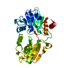
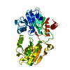
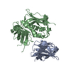
- PDB-5q20: PanDDA analysis group deposition -- Crystal Structure of DCLRE1A ... -
+ Open data

- Basic information
| Entry | Database: PDB / ID: 5q20 | ||||||
|---|---|---|---|---|---|---|---|
| Title | PanDDA analysis group deposition -- Crystal Structure of DCLRE1A in complex with FMOPL000524a | ||||||
 Components Components | DNA cross-link repair 1A protein | ||||||
 Keywords Keywords |  CELL CYCLE / SGC - Diamond I04-1 fragment screening / PanDDA / XChemExplorer CELL CYCLE / SGC - Diamond I04-1 fragment screening / PanDDA / XChemExplorer | ||||||
| Function / homology |  Function and homology information Function and homology information5'-3' DNA exonuclease activity / interstrand cross-link repair / Fanconi Anemia Pathway / fibrillar center / beta-lactamase activity / double-strand break repair via nonhomologous end joining / beta-lactamase / damaged DNA binding / cell cycle / cell division ...5'-3' DNA exonuclease activity / interstrand cross-link repair / Fanconi Anemia Pathway / fibrillar center / beta-lactamase activity / double-strand break repair via nonhomologous end joining / beta-lactamase / damaged DNA binding / cell cycle / cell division / nucleoplasm / metal ion bindingSimilarity search - Function | ||||||
| Biological species |  Homo sapiens (human) Homo sapiens (human) | ||||||
| Method | X-RAY DIFFRACTION / SYNCHROTRON / FOURIER SYNTHESIS / molecular replacement / Resolution: 1.76 Å | ||||||
 Authors Authors | Newman, J.A. / Aitkenhead, H. / Lee, S.Y. / Kupinska, K. / Burgess-Brown, N. / Tallon, R. / Krojer, T. / von Delft, F. / Arrowsmith, C.H. / Edwards, A. ...Newman, J.A. / Aitkenhead, H. / Lee, S.Y. / Kupinska, K. / Burgess-Brown, N. / Tallon, R. / Krojer, T. / von Delft, F. / Arrowsmith, C.H. / Edwards, A. / Bountra, C. / Gileadi, O. | ||||||
 Citation Citation | Journal: To Be Published Title: PanDDA analysis group deposition Authors: Newman, J.A. / Aitkenhead, H. / Lee, S.Y. / Kupinska, K. / Burgess-Brown, N. / Tallon, R. / Krojer, T. / von Delft, F. / Arrowsmith, C.H. / Edwards, A. / Bountra, C. / Gileadi, O. | ||||||
| History |
|
- Structure visualization
| Structure viewer | Molecule: MolmilJmol/JSmol |
|---|
- Downloads & links
-Download
| PDBx/mmCIF format | 5q20.cif.gz | 90.7 KB | Display | PDBx/mmCIF format |
|---|---|---|---|---|
| PDB format | pdb5q20.ent.gz | 66.9 KB | Display | PDB format |
| PDBx/mmJSON format | 5q20.json.gz | Tree view | PDBx/mmJSON format | |
| Others |  Other downloads Other downloads |
-Validation report
| Arichive directory | https://data.pdbj.org/pub/pdb/validation_reports/q2/5q20ftp://data.pdbj.org/pub/pdb/validation_reports/q2/5q20 | HTTPS FTP |
|---|
-Group deposition
| ID | G_1002034 (26 entries) |
|---|---|
| Title | PanDDA analysis group deposition |
| Type | undefined |
| Description | Nuclease domain of human DCLRE1A screened against the DSPL Fragment Library by X-ray Crystallography at the XChem facility of Diamond Light Source beamline I04-1 |


-Related structure data
| Related structure data |  5ahoS S: Starting model for refinement |
|---|---|
| Similar structure data |


-Links
 PDBj
PDBj
- Assembly
| Deposited unit | 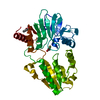
| ||||||||
|---|---|---|---|---|---|---|---|---|---|
| 1 |
| ||||||||
| Unit cell |
|
-Components
| #1: Protein | Mass: 38922.070 Da / Num. of mol.: 1 / Fragment: UNP residues 698-1040 Source method: isolated from a genetically manipulated source Source: (gene. exp.) Homo sapiens (human) / Gene: DCLRE1A, KIAA0086, SNM1, SNM1A / Production host:   Spodoptera frugiperda (fall armyworm) / References: UniProt: Q6PJP8 Spodoptera frugiperda (fall armyworm) / References: UniProt: Q6PJP8 |
|---|---|
| #2: Chemical | ChemComp-MLI / Malonic acid  Mass: 102.046 Da / Num. of mol.: 1 / Source method: obtained synthetically / Formula: C3H2O4 Mass: 102.046 Da / Num. of mol.: 1 / Source method: obtained synthetically / Formula: C3H2O4 |
| #3: Chemical | ChemComp-NI / Nickel  Mass: 58.693 Da / Num. of mol.: 1 / Source method: obtained synthetically / Formula: Ni Mass: 58.693 Da / Num. of mol.: 1 / Source method: obtained synthetically / Formula: Ni |
| #4: Chemical | ChemComp-AYV /   Mass: 224.256 Da / Num. of mol.: 1 / Source method: obtained synthetically / Formula: C11H16N2O3 Mass: 224.256 Da / Num. of mol.: 1 / Source method: obtained synthetically / Formula: C11H16N2O3 |
| #5: Water | ChemComp-HOH / Water Mass: 18.015 Da / Num. of mol.: 306 / Source method: isolated from a natural source / Formula: H2O Mass: 18.015 Da / Num. of mol.: 306 / Source method: isolated from a natural source / Formula: H2O |
| Nonpolymer details | yes CC(CO)(CO)NC(=O)Nc1ccccc1 None -12.9579483987 14.3555341045 14.3555341045 CC(CO)(CO)NC(=O) ... |
-Experimental details
-Experiment
| Experiment | Method: X-RAY DIFFRACTION / Number of used crystals: 1 |
|---|
- Sample preparation
| Crystal | Density Matthews: 2.18 Å3/Da / Density % sol: 43.53 % / Mosaicity: 0 ° |
|---|---|
| Crystal grow | Temperature: 298 K / Method: vapor diffusion, sitting drop / pH: 6 / Details: 30% PEG1000, 0.1 M MIB buffer |
-Data collection
| Diffraction source | Source: SYNCHROTRON / Site: Diamond  / Beamline: I04-1 / Wavelength: 0.92 Å / Beamline: I04-1 / Wavelength: 0.92 Å | |||||||||||||||||||||||||||
|---|---|---|---|---|---|---|---|---|---|---|---|---|---|---|---|---|---|---|---|---|---|---|---|---|---|---|---|---|
| Detector | Type: DECTRIS PILATUS 6M / Detector: PIXEL / Date: Apr 11, 2017 | |||||||||||||||||||||||||||
| Radiation | Monochromator: Si(111) / Protocol: SINGLE WAVELENGTH / Monochromatic (M) / Laue (L): M / Scattering type: x-ray | |||||||||||||||||||||||||||
| Radiation wavelength | Wavelength: 0.92 Å / Relative weight: 1 | |||||||||||||||||||||||||||
| Reflection | Resolution: 1.76→56.82 Å / Num. obs: 34543 / % possible obs: 100 % / Redundancy: 6.3 % / CC1/2: 0.998 / Rmerge(I) obs: 0.111 / Rpim(I) all: 0.048 / Rrim(I) all: 0.121 / Net I/σ(I): 8.5 / Num. measured all: 218723 / Scaling rejects: 24 | |||||||||||||||||||||||||||
| Reflection shell | Diffraction-ID: 1 / Redundancy: 6 %
|
-Phasing
| Phasing | Method: molecular replacement |
|---|
- Processing
| Software |
| |||||||||||||||||||||||||||||||||||||||||||||||||||||||||||||||||||||||||||
|---|---|---|---|---|---|---|---|---|---|---|---|---|---|---|---|---|---|---|---|---|---|---|---|---|---|---|---|---|---|---|---|---|---|---|---|---|---|---|---|---|---|---|---|---|---|---|---|---|---|---|---|---|---|---|---|---|---|---|---|---|---|---|---|---|---|---|---|---|---|---|---|---|---|---|---|---|
| Refinement | Method to determine structure: FOURIER SYNTHESIS Starting model: 5aho Resolution: 1.76→56.82 Å / Cor.coef. Fo:Fc: 0.956 / Cor.coef. Fo:Fc free: 0.934 / SU B: 4.264 / SU ML: 0.123 / Cross valid method: THROUGHOUT / σ(F): 0 / ESU R: 0.143 / ESU R Free: 0.136 / Stereochemistry target values: MAXIMUM LIKELIHOOD Details: HYDROGENS HAVE BEEN ADDED IN THE RIDING POSITIONS U VALUES : REFINED INDIVIDUALLY
| |||||||||||||||||||||||||||||||||||||||||||||||||||||||||||||||||||||||||||
| Solvent computation | Ion probe radii: 0.8 Å / Shrinkage radii: 0.8 Å / VDW probe radii: 1.2 Å / Solvent model: MASK | |||||||||||||||||||||||||||||||||||||||||||||||||||||||||||||||||||||||||||
| Displacement parameters | Biso max: 132.17 Å2 / Biso mean: 26.101 Å2 / Biso min: 8.59 Å2
| |||||||||||||||||||||||||||||||||||||||||||||||||||||||||||||||||||||||||||
| Refinement step | Cycle: final / Resolution: 1.76→56.82 Å
| |||||||||||||||||||||||||||||||||||||||||||||||||||||||||||||||||||||||||||
| Refine LS restraints |
| |||||||||||||||||||||||||||||||||||||||||||||||||||||||||||||||||||||||||||
| LS refinement shell | Resolution: 1.76→1.805 Å / Total num. of bins used: 20
|
