 Movie
Movie Controller


+ Open data

- Basic information
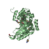
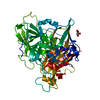
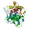
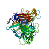
| Entry | Database: PDB / ID: 5oxl | |||||||||
|---|---|---|---|---|---|---|---|---|---|---|
| Title | PepTSt in complex with dipeptide Ala-Leu | |||||||||
 Components Components | Di-or tripeptide:H+ symporter | |||||||||
 Keywords Keywords |  TRANSPORT PROTEIN / Alpha-helical membrane protein / MFS fold / membrane protein / peptide transporter TRANSPORT PROTEIN / Alpha-helical membrane protein / MFS fold / membrane protein / peptide transporter | |||||||||
| Function / homology |  Function and homology information Function and homology informationoligopeptide transport / peptide transmembrane transporter activity / identical protein binding / plasma membraneSimilarity search - Function | |||||||||
| Biological species |  Streptococcus thermophilus (bacteria) Streptococcus thermophilus (bacteria) | |||||||||
| Method | X-RAY DIFFRACTION / SYNCHROTRON / MOLECULAR REPLACEMENT / Resolution: 2.66 Å | |||||||||
 Authors Authors | Martinez Molledo, M. / Quistgaard, E.M. / Loew, C. | |||||||||
 Citation Citation | Journal: Structure / Year: 2018 Title: Multispecific Substrate Recognition in a Proton-Dependent Oligopeptide Transporter. Authors: Martinez Molledo, M. / Quistgaard, E.M. / Flayhan, A. / Pieprzyk, J. / Low, C. | |||||||||
| History |
|
- Structure visualization
| Structure viewer | Molecule: MolmilJmol/JSmol |
|---|
- Downloads & links
-Download
| PDBx/mmCIF format | 5oxl.cif.gz | 195 KB | Display | PDBx/mmCIF format |
|---|---|---|---|---|
| PDB format | pdb5oxl.ent.gz | 156 KB | Display | PDB format |
| PDBx/mmJSON format | 5oxl.json.gz | Tree view | PDBx/mmJSON format | |
| Others |  Other downloads Other downloads |
-Validation report
| Arichive directory | https://data.pdbj.org/pub/pdb/validation_reports/ox/5oxlftp://data.pdbj.org/pub/pdb/validation_reports/ox/5oxl | HTTPS FTP |
|---|
-Related structure data
| Related structure data | 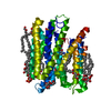 5oxkC 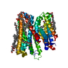 5oxmC 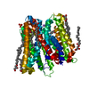 5oxnC 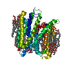 5oxoC 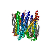 5oxpC 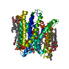 5oxqC 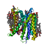 6eiaC 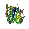 4d2bS S: Starting model for refinement C: citing same article ( |
|---|---|
| Similar structure data |


-Links
 PDBj
PDBj


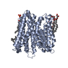
- Assembly
| Deposited unit | 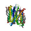
| ||||||||
|---|---|---|---|---|---|---|---|---|---|
| 1 |
| ||||||||
| Unit cell |
|
-Components
-Protein , 1 types, 1 molecules A
| #1: Protein | Mass: 53648.074 Da / Num. of mol.: 1 Source method: isolated from a genetically manipulated source Source: (gene. exp.) Streptococcus thermophilus (strain ATCC BAA-250 / LMG 18311) (bacteria)Strain: ATCC BAA-250 / LMG 18311 / Gene: dtpT, stu0970 / Production host: Escherichia coli (E. coli) / Variant (production host): C41 / References: UniProt: Q5M4H8 |
|---|
-Non-polymers , 8 types, 49 molecules 


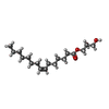
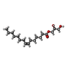


| #2: Chemical | Phosphate Mass: 94.971 Da / Num. of mol.: 2 / Source method: obtained synthetically / Formula: PO4 Mass: 94.971 Da / Num. of mol.: 2 / Source method: obtained synthetically / Formula: PO4#3: Chemical | ChemComp-EPE / | HEPES Mass: 238.305 Da / Num. of mol.: 1 / Source method: obtained synthetically / Formula: C8H18N2O4S / Comment: pH buffer*YM Mass: 238.305 Da / Num. of mol.: 1 / Source method: obtained synthetically / Formula: C8H18N2O4S / Comment: pH buffer*YM#4: Chemical | Polyethylene glycol Mass: 238.278 Da / Num. of mol.: 2 / Source method: obtained synthetically / Formula: C10H22O6 / Comment: precipitant*YM Mass: 238.278 Da / Num. of mol.: 2 / Source method: obtained synthetically / Formula: C10H22O6 / Comment: precipitant*YM#5: Chemical | ChemComp-78N / (  Mass: 314.460 Da / Num. of mol.: 9 / Source method: obtained synthetically / Formula: C18H34O4 Mass: 314.460 Da / Num. of mol.: 9 / Source method: obtained synthetically / Formula: C18H34O4#6: Chemical | ChemComp-78M / ( |  Mass: 314.460 Da / Num. of mol.: 1 / Source method: obtained synthetically / Formula: C18H34O4 Mass: 314.460 Da / Num. of mol.: 1 / Source method: obtained synthetically / Formula: C18H34O4#7: Chemical | ChemComp-ALA / | Alanine Type: L-peptide linking / Mass: 89.093 Da / Num. of mol.: 1 / Source method: obtained synthetically / Formula: C3H7NO2 Type: L-peptide linking / Mass: 89.093 Da / Num. of mol.: 1 / Source method: obtained synthetically / Formula: C3H7NO2#8: Chemical | ChemComp-LEU / | Leucine Type: L-peptide linking / Mass: 131.173 Da / Num. of mol.: 1 / Source method: obtained synthetically / Formula: C6H13NO2 Type: L-peptide linking / Mass: 131.173 Da / Num. of mol.: 1 / Source method: obtained synthetically / Formula: C6H13NO2#9: Water | ChemComp-HOH / | WaterMass: 18.015 Da / Num. of mol.: 32 / Source method: isolated from a natural source / Formula: H2O |
|---|
-Experimental details
-Experiment
| Experiment | Method: X-RAY DIFFRACTION / Number of used crystals: 1 |
|---|
- Sample preparation
| Crystal | Density Matthews: 2.85 Å3/Da / Density % sol: 56.84 % |
|---|---|
| Crystal grow | Temperature: 292.15 K / Method: lipidic cubic phase Details: 0.1 M HEPES pH 7.0, 0.15 M ammonium phosphate monobasic, 18% (v/v) PEG 400, 5 mM Ala-Leu |
-Data collection
| Diffraction | Mean temperature: 100 K |
|---|---|
| Diffraction source | Source: SYNCHROTRON / Site: ESRF  / Beamline: MASSIF-1 / Wavelength: 0.966 Å / Beamline: MASSIF-1 / Wavelength: 0.966 Å |
| Detector | Type: DECTRIS PILATUS3 2M / Detector: PIXEL / Date: Dec 8, 2015 |
| Radiation | Protocol: SINGLE WAVELENGTH / Monochromatic (M) / Laue (L): M / Scattering type: x-ray |
| Radiation wavelength | Wavelength: 0.966 Å / Relative weight: 1 |
| Reflection | Resolution: 2.66→46.27 Å / Num. obs: 19071 / % possible obs: 98.2 % / Redundancy: 3 % / Biso Wilson estimate: 49.82 Å2 / CC1/2: 0.994 / Rmerge(I) obs: 0.1076 / Net I/σ(I): 9.58 |
| Reflection shell | Resolution: 2.66→2.75 Å / Redundancy: 2.9 % / Mean I/σ(I) obs: 1.97 / Num. unique obs: 2881 / CC1/2: 0.72 / % possible all: 96.4 |
- Processing
| Software |
| ||||||||||||||||||||||||||||||||||||||||||||||||||||||||||||||||||||||||||||||||||||||||||||||||||||||||||||||||||||||||||||||||||||||||||||||||||||||||||||||||||||||||||||||||||||||||||||||||||||||||
|---|---|---|---|---|---|---|---|---|---|---|---|---|---|---|---|---|---|---|---|---|---|---|---|---|---|---|---|---|---|---|---|---|---|---|---|---|---|---|---|---|---|---|---|---|---|---|---|---|---|---|---|---|---|---|---|---|---|---|---|---|---|---|---|---|---|---|---|---|---|---|---|---|---|---|---|---|---|---|---|---|---|---|---|---|---|---|---|---|---|---|---|---|---|---|---|---|---|---|---|---|---|---|---|---|---|---|---|---|---|---|---|---|---|---|---|---|---|---|---|---|---|---|---|---|---|---|---|---|---|---|---|---|---|---|---|---|---|---|---|---|---|---|---|---|---|---|---|---|---|---|---|---|---|---|---|---|---|---|---|---|---|---|---|---|---|---|---|---|---|---|---|---|---|---|---|---|---|---|---|---|---|---|---|---|---|---|---|---|---|---|---|---|---|---|---|---|---|---|---|---|---|
| Refinement | Method to determine structure: MOLECULAR REPLACEMENT Starting model: 4D2B Resolution: 2.66→46.266 Å / SU ML: 0.31 / Cross valid method: FREE R-VALUE / Phase error: 24.73
| ||||||||||||||||||||||||||||||||||||||||||||||||||||||||||||||||||||||||||||||||||||||||||||||||||||||||||||||||||||||||||||||||||||||||||||||||||||||||||||||||||||||||||||||||||||||||||||||||||||||||
| Solvent computation | Shrinkage radii: 0.9 Å / VDW probe radii: 1.11 Å | ||||||||||||||||||||||||||||||||||||||||||||||||||||||||||||||||||||||||||||||||||||||||||||||||||||||||||||||||||||||||||||||||||||||||||||||||||||||||||||||||||||||||||||||||||||||||||||||||||||||||
| Refinement step | Cycle: LAST / Resolution: 2.66→46.266 Å
| ||||||||||||||||||||||||||||||||||||||||||||||||||||||||||||||||||||||||||||||||||||||||||||||||||||||||||||||||||||||||||||||||||||||||||||||||||||||||||||||||||||||||||||||||||||||||||||||||||||||||
| Refine LS restraints |
| ||||||||||||||||||||||||||||||||||||||||||||||||||||||||||||||||||||||||||||||||||||||||||||||||||||||||||||||||||||||||||||||||||||||||||||||||||||||||||||||||||||||||||||||||||||||||||||||||||||||||
| LS refinement shell |
| ||||||||||||||||||||||||||||||||||||||||||||||||||||||||||||||||||||||||||||||||||||||||||||||||||||||||||||||||||||||||||||||||||||||||||||||||||||||||||||||||||||||||||||||||||||||||||||||||||||||||
| Refinement TLS params. | Method: refined / Refine-ID: X-RAY DIFFRACTION
| ||||||||||||||||||||||||||||||||||||||||||||||||||||||||||||||||||||||||||||||||||||||||||||||||||||||||||||||||||||||||||||||||||||||||||||||||||||||||||||||||||||||||||||||||||||||||||||||||||||||||
| Refinement TLS group |
|
