 Movie
Movie Controller

[English] 日本語
 Yorodumi
- PDB-5out: CRYSTAL STRUCTURE OF THE FERRIC ENTEROBACTIN RECEPTOR (PFEA) MUTA... -
+ Open data

- Basic information
| Entry | Database: PDB / ID: 5out | ||||||
|---|---|---|---|---|---|---|---|
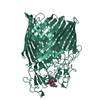
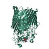
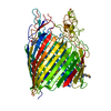
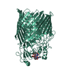
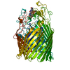
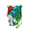
| Title | CRYSTAL STRUCTURE OF THE FERRIC ENTEROBACTIN RECEPTOR (PFEA) MUTANT (G324V) FROM PSEUDOMONAS AERUGINOSA | ||||||
 Components Components | Ferric enterobactin receptor | ||||||
 Keywords Keywords |  MEMBRANE PROTEIN / PfeA / TBDT MEMBRANE PROTEIN / PfeA / TBDT | ||||||
| Function / homology |  Function and homology information Function and homology informationenterobactin transmembrane transporter activity / enterobactin transport / colicin transmembrane transporter activity / siderophore transmembrane transport / siderophore uptake transmembrane transporter activity / siderophore transport / outer membrane / enterobactin binding / cell outer membrane / signaling receptor activitySimilarity search - Function | ||||||
| Biological species |  Pseudomonas aeruginosa PAO1 (bacteria) Pseudomonas aeruginosa PAO1 (bacteria) | ||||||
| Method | X-RAY DIFFRACTION / Resolution: 2.9 Å | ||||||
 Authors Authors | Moynie, L. / Naismith, J.H. | ||||||
 Citation Citation | Journal: Nat Commun / Year: 2019 Title: The complex of ferric-enterobactin with its transporter from Pseudomonas aeruginosa suggests a two-site model. Authors: Moynie, L. / Milenkovic, S. / Mislin, G.L.A. / Gasser, V. / Malloci, G. / Baco, E. / McCaughan, R.P. / Page, M.G.P. / Schalk, I.J. / Ceccarelli, M. / Naismith, J.H. | ||||||
| History |
|
- Structure visualization
| Structure viewer | Molecule: MolmilJmol/JSmol |
|---|
- Downloads & links
-Download
| PDBx/mmCIF format | 5out.cif.gz | 279.2 KB | Display | PDBx/mmCIF format |
|---|---|---|---|---|
| PDB format | pdb5out.ent.gz | 235.9 KB | Display | PDB format |
| PDBx/mmJSON format | 5out.json.gz | Tree view | PDBx/mmJSON format | |
| Others |  Other downloads Other downloads |
-Validation report
| Arichive directory | https://data.pdbj.org/pub/pdb/validation_reports/ou/5outftp://data.pdbj.org/pub/pdb/validation_reports/ou/5out | HTTPS FTP |
|---|
-Related structure data
| Related structure data | 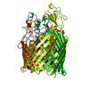 5m9bC 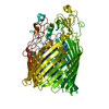 5mzsC  5nc4C  5nr2C  6i2jC 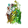 6q5eC  6r1fC C: citing same article ( |
|---|---|
| Similar structure data |


-Links
 PDBj
PDBj- Assembly
| Deposited unit | 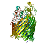
| ||||||||
|---|---|---|---|---|---|---|---|---|---|
| 1 |
| ||||||||
| Unit cell |
|
-Components
| #1: Protein | Mass: 78527.203 Da / Num. of mol.: 1 / Mutation: G324V Source method: isolated from a genetically manipulated source Source: (gene. exp.) Pseudomonas aeruginosa PAO1 (bacteria) / Gene: pfeA, PA2688 / Production host: Escherichia coli (E. coli) / References: UniProt: Q05098 |
|---|---|
| #2: Water | ChemComp-HOH / Water Mass: 18.015 Da / Num. of mol.: 7 / Source method: isolated from a natural source / Formula: H2O Mass: 18.015 Da / Num. of mol.: 7 / Source method: isolated from a natural source / Formula: H2O |
-Experimental details
-Experiment
| Experiment | Method: X-RAY DIFFRACTION / Number of used crystals: 1 |
|---|
- Sample preparation
| Crystal | Density Matthews: 3.44 Å3/Da / Density % sol: 64.22 % |
|---|---|
| Crystal grow | Temperature: 294 K / Method: vapor diffusion / Details: PEG 8000 ADA Magnesium acetate pH 6.5 |
-Data collection
| Diffraction | Mean temperature: 100 K |
|---|---|
| Diffraction source | Source: ROTATING ANODE / Type: RIGAKU MICROMAX-007 HF / Wavelength: 1.54178 Å |
| Detector | Type: RIGAKU SATURN 944+ / Detector: CCD / Date: Aug 26, 2016 |
| Radiation | Protocol: SINGLE WAVELENGTH / Monochromatic (M) / Laue (L): M / Scattering type: x-ray |
| Radiation wavelength | Wavelength: 1.54178 Å / Relative weight: 1 |
| Reflection | Resolution: 2.9→39.08 Å / Num. obs: 19876 / % possible obs: 80.4 % / Redundancy: 3.5 % / CC1/2: 0.998 / Rmerge(I) obs: 0.106 / Net I/σ(I): 12.7 |
| Reflection shell | Resolution: 2.9→2.95 Å / Redundancy: 3.5 % / Rmerge(I) obs: 1.377 / Mean I/σ(I) obs: 1.4 / CC1/2: 0.669 / % possible all: 81.9 |
- Processing
| Software |
| ||||||||||||||||||||||||||||||||||||||||||||||||||||||||||||||||||||||||||||||||||||||||||||||||||||||||||||||||||||||||||||||||||||||||||||||||||||||||||||||||||||||||||||||||||||||
|---|---|---|---|---|---|---|---|---|---|---|---|---|---|---|---|---|---|---|---|---|---|---|---|---|---|---|---|---|---|---|---|---|---|---|---|---|---|---|---|---|---|---|---|---|---|---|---|---|---|---|---|---|---|---|---|---|---|---|---|---|---|---|---|---|---|---|---|---|---|---|---|---|---|---|---|---|---|---|---|---|---|---|---|---|---|---|---|---|---|---|---|---|---|---|---|---|---|---|---|---|---|---|---|---|---|---|---|---|---|---|---|---|---|---|---|---|---|---|---|---|---|---|---|---|---|---|---|---|---|---|---|---|---|---|---|---|---|---|---|---|---|---|---|---|---|---|---|---|---|---|---|---|---|---|---|---|---|---|---|---|---|---|---|---|---|---|---|---|---|---|---|---|---|---|---|---|---|---|---|---|---|---|---|
| Refinement | Resolution: 2.9→39.08 Å / Cor.coef. Fo:Fc: 0.898 / Cor.coef. Fo:Fc free: 0.88 / SU B: 57.47 / SU ML: 0.423 / Cross valid method: THROUGHOUT / ESU R Free: 0.505 / Details: HYDROGENS HAVE BEEN ADDED IN THE RIDING POSITIONS
| ||||||||||||||||||||||||||||||||||||||||||||||||||||||||||||||||||||||||||||||||||||||||||||||||||||||||||||||||||||||||||||||||||||||||||||||||||||||||||||||||||||||||||||||||||||||
| Solvent computation | Ion probe radii: 0.8 Å / Shrinkage radii: 0.8 Å / VDW probe radii: 1.1 Å | ||||||||||||||||||||||||||||||||||||||||||||||||||||||||||||||||||||||||||||||||||||||||||||||||||||||||||||||||||||||||||||||||||||||||||||||||||||||||||||||||||||||||||||||||||||||
| Displacement parameters | Biso mean: 69.496 Å2
| ||||||||||||||||||||||||||||||||||||||||||||||||||||||||||||||||||||||||||||||||||||||||||||||||||||||||||||||||||||||||||||||||||||||||||||||||||||||||||||||||||||||||||||||||||||||
| Refinement step | Cycle: 1 / Resolution: 2.9→39.08 Å
| ||||||||||||||||||||||||||||||||||||||||||||||||||||||||||||||||||||||||||||||||||||||||||||||||||||||||||||||||||||||||||||||||||||||||||||||||||||||||||||||||||||||||||||||||||||||
| Refine LS restraints |
|