 Movie
Movie Controller

[English] 日本語
 Yorodumi
- PDB-5or6: Crystal structures of PYR1/HAB1 in complex with synthetic analogu... -
+ Open data

- Basic information
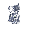
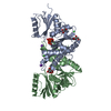
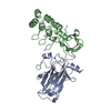
| Entry | Database: PDB / ID: 5or6 | ||||||
|---|---|---|---|---|---|---|---|
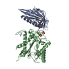
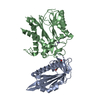
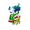
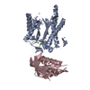
| Title | Crystal structures of PYR1/HAB1 in complex with synthetic analogues of Abscisic Acid | ||||||
 Components Components |
| ||||||
 Keywords Keywords |  PROTEIN BINDING / Complex Arabidopsis hab1-pyr1 PROTEIN BINDING / Complex Arabidopsis hab1-pyr1 | ||||||
| Function / homology |  Function and homology information Function and homology informationpositive regulation of response to water deprivation / regulation of protein serine/threonine phosphatase activity / plant-type vacuole membrane / protein phosphatase inhibitor complex / abscisic acid binding / abscisic acid-activated signaling pathway / protein phosphatase inhibitor activity / myosin phosphatase activity / protein serine/threonine phosphatase activity / protein-serine/threonine phosphatase ...positive regulation of response to water deprivation / regulation of protein serine/threonine phosphatase activity / plant-type vacuole membrane / protein phosphatase inhibitor complex / abscisic acid binding / abscisic acid-activated signaling pathway / protein phosphatase inhibitor activity / myosin phosphatase activity / protein serine/threonine phosphatase activity / protein-serine/threonine phosphatase / ubiquitin-like protein ligase binding / signaling receptor activity / protein homodimerization activity / identical protein binding / metal ion binding / nucleus / plasma membrane / cytosol / cytoplasmSimilarity search - Function | ||||||
| Biological species |  Arabidopsis thaliana (thale cress) Arabidopsis thaliana (thale cress) | ||||||
| Method | X-RAY DIFFRACTION / MOLECULAR REPLACEMENT / Resolution: 2.4 Å | ||||||
 Authors Authors | Freigang, J. | ||||||
 Citation Citation | Journal: Eur.J.Org.Chem. / Year: 2018 Title: Insights into the in Vitro and in Vivo SAR of Abscisic Acid - Exploring Unprecedented Variations of the Side Chain via Cross-Coupling-Mediated Syntheses Authors: Frackenpohl, J. / Grill, E. / Bojack, G. / Baltz, R. / Busch, M. / Dittgen, J. / Franke, J. / Freigang, J. / Gonzalez, S. / Heinemann, I. / Helmke, H. / Hills, M. / Hohmann, S. / von Koskull- ...Authors: Frackenpohl, J. / Grill, E. / Bojack, G. / Baltz, R. / Busch, M. / Dittgen, J. / Franke, J. / Freigang, J. / Gonzalez, S. / Heinemann, I. / Helmke, H. / Hills, M. / Hohmann, S. / von Koskull-Doering, P. / Kleemann, J. / Lange, G. / Lehr, S. / Mueller, T. / Peschel, E. / Poree, F. / Schmutzler, D. / Schulz, A. / Willms, L. / Wunschel, C. | ||||||
| History |
|
- Structure visualization
| Structure viewer | Molecule: MolmilJmol/JSmol |
|---|
- Downloads & links
-Download
| PDBx/mmCIF format | 5or6.cif.gz | 121.2 KB | Display | PDBx/mmCIF format |
|---|---|---|---|---|
| PDB format | pdb5or6.ent.gz | 88.6 KB | Display | PDB format |
| PDBx/mmJSON format | 5or6.json.gz | Tree view | PDBx/mmJSON format | |
| Others |  Other downloads Other downloads |
-Validation report
| Arichive directory | https://data.pdbj.org/pub/pdb/validation_reports/or/5or6ftp://data.pdbj.org/pub/pdb/validation_reports/or/5or6 | HTTPS FTP |
|---|
-Related structure data
| Related structure data |  5or2C  3qn1S C: citing same article ( S: Starting model for refinement |
|---|---|
| Similar structure data |


-Links
 PDBj
PDBj- Assembly
| Deposited unit | 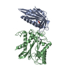
| ||||||||
|---|---|---|---|---|---|---|---|---|---|
| 1 |
| ||||||||
| Unit cell |
|
-Components
| #1: Protein | Mass: 21705.340 Da / Num. of mol.: 1 Source method: isolated from a genetically manipulated source Source: (gene. exp.) Arabidopsis thaliana (thale cress) / Gene: PYR1, ABIP6, RCAR11, At4g17870, T6K21.50 / Production host:  Escherichia coli (E. coli) / References: UniProt: O49686 Escherichia coli (E. coli) / References: UniProt: O49686 | ||
|---|---|---|---|
| #2: Protein | Mass: 37347.824 Da / Num. of mol.: 1 Source method: isolated from a genetically manipulated source Source: (gene. exp.) Arabidopsis thaliana (thale cress) / Gene: HAB1, P2C-HA, At1g72770, F28P22.4 / Production host: Escherichia coli (E. coli)References: UniProt: Q9CAJ0, protein-serine/threonine phosphatase | ||
| #3: Chemical | ChemComp-A4K / (~{  Mass: 316.272 Da / Num. of mol.: 1 / Source method: obtained synthetically / Formula: C15H15F3O4 / Feature type: SUBJECT OF INVESTIGATION Mass: 316.272 Da / Num. of mol.: 1 / Source method: obtained synthetically / Formula: C15H15F3O4 / Feature type: SUBJECT OF INVESTIGATION | ||
| #4: Chemical |   Mass: 54.938 Da / Num. of mol.: 3 / Source method: isolated from a natural source / Formula: Mn Mass: 54.938 Da / Num. of mol.: 3 / Source method: isolated from a natural source / Formula: Mn#5: Water | ChemComp-HOH / | Water Mass: 18.015 Da / Num. of mol.: 465 / Source method: isolated from a natural source / Formula: H2O Mass: 18.015 Da / Num. of mol.: 465 / Source method: isolated from a natural source / Formula: H2O |
-Experimental details
-Experiment
| Experiment | Method: X-RAY DIFFRACTION / Number of used crystals: 1 |
|---|
- Sample preparation
| Crystal | Density Matthews: 2.33 Å3/Da / Density % sol: 47.25 % |
|---|---|
| Crystal grow | Temperature: 278 K / Method: vapor diffusion, sitting drop Details: 20% Peg 8000, 100 mM Tris pH 8.5, 160 mM MgCl2, 60 mM glycylglycylglycine |
-Data collection
| Diffraction | Mean temperature: 100 K |
|---|---|
| Diffraction source | Source: SEALED TUBE / Type: OXFORD DIFFRACTION NOVA / Wavelength: 1.54 Å |
| Detector | Type: OXFORD ONYX CCD / Detector: CCD / Date: Apr 3, 2012 |
| Radiation | Protocol: SINGLE WAVELENGTH / Monochromatic (M) / Laue (L): M / Scattering type: x-ray |
| Radiation wavelength | Wavelength: 1.54 Å / Relative weight: 1 |
| Reflection | Resolution: 2.4→20 Å / Num. obs: 20069 / % possible obs: 97 % / Redundancy: 3.4 % / Rsym value: 0.1 / Net I/σ(I): 10.53 |
| Reflection shell | Resolution: 2.4→2.55 Å / Rsym value: 0.351 |
- Processing
| Software |
| ||||||||||||||||||||||||||||||||||||||||||||||||||||||||||||||||||||||||||||||||||||||||||||||||||||||||||||||||||||||||||||||||||||||||||||||||||||||||||||||||||||||||||||||||||||||
|---|---|---|---|---|---|---|---|---|---|---|---|---|---|---|---|---|---|---|---|---|---|---|---|---|---|---|---|---|---|---|---|---|---|---|---|---|---|---|---|---|---|---|---|---|---|---|---|---|---|---|---|---|---|---|---|---|---|---|---|---|---|---|---|---|---|---|---|---|---|---|---|---|---|---|---|---|---|---|---|---|---|---|---|---|---|---|---|---|---|---|---|---|---|---|---|---|---|---|---|---|---|---|---|---|---|---|---|---|---|---|---|---|---|---|---|---|---|---|---|---|---|---|---|---|---|---|---|---|---|---|---|---|---|---|---|---|---|---|---|---|---|---|---|---|---|---|---|---|---|---|---|---|---|---|---|---|---|---|---|---|---|---|---|---|---|---|---|---|---|---|---|---|---|---|---|---|---|---|---|---|---|---|---|
| Refinement | Method to determine structure: MOLECULAR REPLACEMENT Starting model: 3QN1 Resolution: 2.4→20 Å / Cor.coef. Fo:Fc: 0.908 / Cor.coef. Fo:Fc free: 0.843 / SU B: 11.599 / SU ML: 0.266 / Cross valid method: THROUGHOUT / ESU R: 0.739 / ESU R Free: 0.337 / Stereochemistry target values: MAXIMUM LIKELIHOOD / Details: HYDROGENS HAVE BEEN ADDED IN THE RIDING POSITIONS
| ||||||||||||||||||||||||||||||||||||||||||||||||||||||||||||||||||||||||||||||||||||||||||||||||||||||||||||||||||||||||||||||||||||||||||||||||||||||||||||||||||||||||||||||||||||||
| Solvent computation | Ion probe radii: 0.8 Å / Shrinkage radii: 0.8 Å / VDW probe radii: 1.4 Å / Solvent model: MASK | ||||||||||||||||||||||||||||||||||||||||||||||||||||||||||||||||||||||||||||||||||||||||||||||||||||||||||||||||||||||||||||||||||||||||||||||||||||||||||||||||||||||||||||||||||||||
| Displacement parameters | Biso mean: 26.881 Å2
| ||||||||||||||||||||||||||||||||||||||||||||||||||||||||||||||||||||||||||||||||||||||||||||||||||||||||||||||||||||||||||||||||||||||||||||||||||||||||||||||||||||||||||||||||||||||
| Refinement step | Cycle: 1 / Resolution: 2.4→20 Å
| ||||||||||||||||||||||||||||||||||||||||||||||||||||||||||||||||||||||||||||||||||||||||||||||||||||||||||||||||||||||||||||||||||||||||||||||||||||||||||||||||||||||||||||||||||||||
| Refine LS restraints |
|