 Movie
Movie Controller


+ Open data

- Basic information
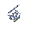
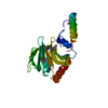
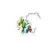
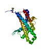
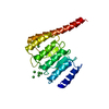
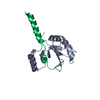
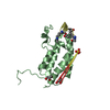
| Entry | Database: PDB / ID: 5ond | |||||||||
|---|---|---|---|---|---|---|---|---|---|---|
| Title | RfaH from Escherichia coli in complex with ops DNA | |||||||||
 Components Components |
| |||||||||
 Keywords Keywords |  TRANSCRIPTION / operon-specific transcription factor / transformer protein / binding of single stranded DNA / translation activation TRANSCRIPTION / operon-specific transcription factor / transformer protein / binding of single stranded DNA / translation activation | |||||||||
| Function / homology |  Function and homology informationregulatory RNA binding / transcription antitermination factor activity, DNA binding / translation activator activity / DNA-templated transcription elongation / bacterial-type RNA polymerase core enzyme binding / transcription elongation-coupled chromatin remodeling / transcription antitermination / positive regulation of translation / DNA binding / cytosol Function and homology informationregulatory RNA binding / transcription antitermination factor activity, DNA binding / translation activator activity / DNA-templated transcription elongation / bacterial-type RNA polymerase core enzyme binding / transcription elongation-coupled chromatin remodeling / transcription antitermination / positive regulation of translation / DNA binding / cytosolSimilarity search - Function | |||||||||
| Biological species |  Escherichia coli (E. coli) Escherichia coli (E. coli) | |||||||||
| Method | X-RAY DIFFRACTION / SYNCHROTRON / MOLECULAR REPLACEMENT / Resolution: 2.1 Å | |||||||||
 Authors Authors | Zuber, P.K. / Artsimovitch, I. / Roesch, P. / Knauer, S.H. | |||||||||
| Funding support |  Germany, 2items Germany, 2items
| |||||||||
 Citation Citation | Journal: Elife / Year: 2018 Title: The universally-conserved transcription factor RfaH is recruited to a hairpin structure of the non-template DNA strand. Authors: Zuber, P.K. / Artsimovitch, I. / NandyMazumdar, M. / Liu, Z. / Nedialkov, Y. / Schweimer, K. / Rosch, P. / Knauer, S.H. | |||||||||
| History |
|
- Structure visualization
| Structure viewer | Molecule: MolmilJmol/JSmol |
|---|
- Downloads & links
-Download
| PDBx/mmCIF format | 5ond.cif.gz | 130.8 KB | Display | PDBx/mmCIF format |
|---|---|---|---|---|
| PDB format | pdb5ond.ent.gz | 101.5 KB | Display | PDB format |
| PDBx/mmJSON format | 5ond.json.gz | Tree view | PDBx/mmJSON format | |
| Others |  Other downloads Other downloads |
-Validation report
| Arichive directory | https://data.pdbj.org/pub/pdb/validation_reports/on/5ondftp://data.pdbj.org/pub/pdb/validation_reports/on/5ond | HTTPS FTP |
|---|
-Related structure data
| Related structure data |  2ougS S: Starting model for refinement |
|---|---|
| Similar structure data |


-Links
 PDBj
PDBj

- Assembly
| Deposited unit | 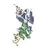
| ||||||||
|---|---|---|---|---|---|---|---|---|---|
| 1 | 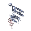
| ||||||||
| 2 | 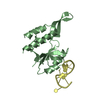
| ||||||||
| Unit cell |
|
-Components
| #1: Protein | Mass: 18322.137 Da / Num. of mol.: 2 / Mutation: L162A Source method: isolated from a genetically manipulated source Source: (gene. exp.) Escherichia coli (E. coli) / Gene: rfaH, hlyT, sfrB, b3842, JW3818 / Production host: Escherichia coli BL21(DE3) (bacteria) / References: UniProt: P0AFW0#2: DNA chain | Mass: 2771.822 Da / Num. of mol.: 2 / Source method: obtained synthetically / Source: (synth.) Escherichia coli (E. coli)#3: Water | ChemComp-HOH / | Water Mass: 18.015 Da / Num. of mol.: 116 / Source method: isolated from a natural source / Formula: H2O Mass: 18.015 Da / Num. of mol.: 116 / Source method: isolated from a natural source / Formula: H2O |
|---|
-Experimental details
-Experiment
| Experiment | Method: X-RAY DIFFRACTION / Number of used crystals: 1 |
|---|
- Sample preparation
| Crystal | Density Matthews: 2.15 Å3/Da / Density % sol: 48.84 % / Description: parallelepipedic, thin |
|---|---|
| Crystal grow | Temperature: 277 K / Method: vapor diffusion, hanging drop / pH: 7.8 Details: protein:DNA molar ratio: 1:1 reservoir solution: 21 %(w/v) PEG monomethyl ether 550, 44.4 mM HEPES (pH 7.0), 4 mM MgCl2; drops: 2ul protein/DNA solution + 2 ul reservoir solution |
-Data collection
| Diffraction | Mean temperature: 100 K |
|---|---|
| Diffraction source | Source: SYNCHROTRON / Site: BESSY / Beamline: 14.1 / Wavelength: 0.9184 Å |
| Detector | Type: DECTRIS PILATUS 6M / Detector: PIXEL / Date: Jan 20, 2017 |
| Radiation | Monochromator: Si-111 crystal / Protocol: SINGLE WAVELENGTH / Monochromatic (M) / Laue (L): M / Scattering type: x-ray |
| Radiation wavelength | Wavelength: 0.9184 Å / Relative weight: 1 |
| Reflection | Resolution: 2.1→41.55 Å / Num. obs: 19931 / % possible obs: 97.3 % / Redundancy: 5.4 % / Rsym value: 0.063 / Net I/σ(I): 13.96 |
| Reflection shell | Resolution: 2.1→2.2 Å / Redundancy: 5.4 % / Mean I/σ(I) obs: 3.47 / Num. unique obs: 2633 / Rsym value: 0.429 / % possible all: 97.9 |
- Processing
| Software |
| ||||||||||||||||||||||||||||||||||||||||||||||||||||||||
|---|---|---|---|---|---|---|---|---|---|---|---|---|---|---|---|---|---|---|---|---|---|---|---|---|---|---|---|---|---|---|---|---|---|---|---|---|---|---|---|---|---|---|---|---|---|---|---|---|---|---|---|---|---|---|---|---|---|
| Refinement | Method to determine structure: MOLECULAR REPLACEMENT Starting model: 2OUG Resolution: 2.1→41.542 Å / SU ML: 0.24 / Cross valid method: FREE R-VALUE / σ(F): 1.98 / Phase error: 32.04 / Stereochemistry target values: ML
| ||||||||||||||||||||||||||||||||||||||||||||||||||||||||
| Solvent computation | Shrinkage radii: 0.9 Å / VDW probe radii: 1.11 Å / Solvent model: FLAT BULK SOLVENT MODEL | ||||||||||||||||||||||||||||||||||||||||||||||||||||||||
| Refinement step | Cycle: LAST / Resolution: 2.1→41.542 Å
| ||||||||||||||||||||||||||||||||||||||||||||||||||||||||
| Refine LS restraints |
| ||||||||||||||||||||||||||||||||||||||||||||||||||||||||
| LS refinement shell |
|
