 Movie
Movie Controller

[English] 日本語
 Yorodumi
- PDB-5og0: Crystal structure of human Alanine:Glyoxylate Aminotransferase ma... -
+ Open data

- Basic information
| Entry | Database: PDB / ID: 5og0 | ||||||
|---|---|---|---|---|---|---|---|
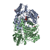
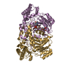
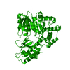
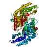
| Title | Crystal structure of human Alanine:Glyoxylate Aminotransferase major allele (AGT-Ma) at 2.5 Angstrom; internal aldimine with PLP in the active site | ||||||
 Components Components | Serine--pyruvate aminotransferase | ||||||
 Keywords Keywords |  TRANSFERASE / AMINOTRANSFERASE / DETOXIFICATION / LIVER TRANSFERASE / AMINOTRANSFERASE / DETOXIFICATION / LIVER | ||||||
| Function / homology |  Function and homology information Function and homology informationoxalic acid secretion / serine-pyruvate transaminase / alanine-glyoxylate transaminase / glycine biosynthetic process, by transamination of glyoxylate / glyoxylate metabolic process / serine-pyruvate transaminase activity / L-alanine catabolic process / alanine-glyoxylate transaminase activity / L-serine metabolic process / L-cysteine catabolic process ...oxalic acid secretion / serine-pyruvate transaminase / alanine-glyoxylate transaminase / glycine biosynthetic process, by transamination of glyoxylate / glyoxylate metabolic process / serine-pyruvate transaminase activity / L-alanine catabolic process / alanine-glyoxylate transaminase activity / L-serine metabolic process / L-cysteine catabolic process / glyoxylate catabolic process / Glyoxylate metabolism and glycine degradation / transaminase activity / amino acid binding / peroxisomal matrix / Notch signaling pathway / Peroxisomal protein import / peroxisome / : / pyridoxal phosphate binding / intracellular membrane-bounded organelle / protein homodimerization activity / identical protein binding / cytosolSimilarity search - Function | ||||||
| Biological species |  Homo sapiens (human) Homo sapiens (human) | ||||||
| Method | X-RAY DIFFRACTION / SYNCHROTRON / MOLECULAR REPLACEMENT / molecular replacement / Resolution: 2.5 Å | ||||||
 Authors Authors | Giardina, G. / Cutruzzola, F. / Borri Voltattorni, C. / Cellini, B. / Montioli, R. | ||||||
 Citation Citation | Journal: Sci Rep / Year: 2017 Title: Radiation damage at the active site of human alanine:glyoxylate aminotransferase reveals that the cofactor position is finely tuned during catalysis. Authors: Giardina, G. / Paiardini, A. / Montioli, R. / Cellini, B. / Voltattorni, C.B. / Cutruzzola, F. | ||||||
| History |
|
- Structure visualization
| Structure viewer | Molecule: MolmilJmol/JSmol |
|---|
- Downloads & links
-Download
| PDBx/mmCIF format | 5og0.cif.gz | 87.7 KB | Display | PDBx/mmCIF format |
|---|---|---|---|---|
| PDB format | pdb5og0.ent.gz | 65.7 KB | Display | PDB format |
| PDBx/mmJSON format | 5og0.json.gz | Tree view | PDBx/mmJSON format | |
| Others |  Other downloads Other downloads |
-Validation report
| Arichive directory | https://data.pdbj.org/pub/pdb/validation_reports/og/5og0ftp://data.pdbj.org/pub/pdb/validation_reports/og/5og0 | HTTPS FTP |
|---|
-Related structure data
| Related structure data |  5f9sSC  5hhyC  5lucC  5ofyC S: Starting model for refinement C: citing same article ( |
|---|---|
| Similar structure data |


-Links
 PDBj
PDBj- Assembly
| Deposited unit | 
| ||||||||
|---|---|---|---|---|---|---|---|---|---|
| 1 | 
| ||||||||
| Unit cell |
|
-Components
| #1: Protein | Mass: 43062.840 Da / Num. of mol.: 1 Source method: isolated from a genetically manipulated source Source: (gene. exp.) Homo sapiens (human) / Gene: AGXT, AGT1, SPAT / Production host:  Escherichia coli (E. coli) Escherichia coli (E. coli)References: UniProt: P21549, serine-pyruvate transaminase, alanine-glyoxylate transaminase |
|---|---|
| #2: Chemical | ChemComp-PLP / Pyridoxal phosphate  Mass: 247.142 Da / Num. of mol.: 1 / Source method: obtained synthetically / Formula: C8H10NO6P Mass: 247.142 Da / Num. of mol.: 1 / Source method: obtained synthetically / Formula: C8H10NO6P |
| #3: Water | ChemComp-HOH / Water Mass: 18.015 Da / Num. of mol.: 43 / Source method: isolated from a natural source / Formula: H2O Mass: 18.015 Da / Num. of mol.: 43 / Source method: isolated from a natural source / Formula: H2O |
-Experimental details
-Experiment
| Experiment | Method: X-RAY DIFFRACTION / Number of used crystals: 1 |
|---|
- Sample preparation
| Crystal | Density Matthews: 3.35 Å3/Da / Density % sol: 63.3 % |
|---|---|
| Crystal grow | Temperature: 294 K / Method: vapor diffusion, hanging drop Details: Protein solution; 0.2 M AGT, 18mM potassium phosphate pH7.4, 20mM Hepes pH 7.4, 5% Jeffamine (Hampton), 5mM sodium hydroxylamine. Reservoir; PEG 6k 12%, 100 mM MES pH 5.0. Mixing: 1+1 microL ...Details: Protein solution; 0.2 M AGT, 18mM potassium phosphate pH7.4, 20mM Hepes pH 7.4, 5% Jeffamine (Hampton), 5mM sodium hydroxylamine. Reservoir; PEG 6k 12%, 100 mM MES pH 5.0. Mixing: 1+1 microL Cryoprotectant; 25% MPD |
-Data collection
| Diffraction | Mean temperature: 100 K | |||||||||||||||||||||
|---|---|---|---|---|---|---|---|---|---|---|---|---|---|---|---|---|---|---|---|---|---|---|
| Diffraction source | Source: SYNCHROTRON / Site: ELETTRA  / Beamline: 5.2R / Wavelength: 1 Å / Beamline: 5.2R / Wavelength: 1 Å | |||||||||||||||||||||
| Detector | Type: DECTRIS PILATUS 2M / Detector: PIXEL / Date: Mar 29, 2015 | |||||||||||||||||||||
| Radiation | Protocol: SINGLE WAVELENGTH / Monochromatic (M) / Laue (L): M / Scattering type: x-ray | |||||||||||||||||||||
| Radiation wavelength | Wavelength: 1 Å / Relative weight: 1 | |||||||||||||||||||||
| Reflection | Resolution: 2.5→47.46 Å / Num. obs: 21107 / % possible obs: 100 % / Redundancy: 17.5 % / CC1/2: 0.999 / Rmerge(I) obs: 0.116 / Rpim(I) all: 0.028 / Rrim(I) all: 0.119 / Net I/σ(I): 19 | |||||||||||||||||||||
| Reflection shell | Diffraction-ID: 1
|
-Phasing
| Phasing | Method: molecular replacement | ||||||
|---|---|---|---|---|---|---|---|
| Phasing MR | R rigid body: 0.41
|
- Processing
| Software |
| ||||||||||||||||||||
|---|---|---|---|---|---|---|---|---|---|---|---|---|---|---|---|---|---|---|---|---|---|
| Refinement | Method to determine structure: MOLECULAR REPLACEMENT Starting model: 5F9S Resolution: 2.5→45 Å / SU B: 13.713 / SU ML: 0.276 / Cross valid method: THROUGHOUT / σ(F): 0 / ESU R: 0.402 / ESU R Free: 0.292 / Details: HYDROGENS HAVE BEEN ADDED IN THE RIDING
| ||||||||||||||||||||
| Displacement parameters | Biso mean: 54.61 Å2
| ||||||||||||||||||||
| Refinement step | Cycle: 1 / Resolution: 2.5→45 Å
|
