 Movie
Movie Controller

[English] 日本語
 Yorodumi
- PDB-5ocp: The periplasmic binding protein component of the arabinose ABC tr... -
+ Open data

- Basic information
| Entry | Database: PDB / ID: 5ocp | ||||||||||||
|---|---|---|---|---|---|---|---|---|---|---|---|---|---|
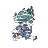
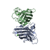
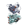
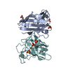
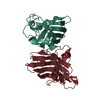
| Title | The periplasmic binding protein component of the arabinose ABC transporter from Shewanella sp. ANA-3 bound to alpha and beta-L-arabinofuranose | ||||||||||||
 Components Components | Periplasmic binding protein/LacI transcriptional regulator | ||||||||||||
 Keywords Keywords | SUGAR BINDING PROTEIN /  ABC transporter / periplasmic / binding protein / arabinose / arabinofuranose ABC transporter / periplasmic / binding protein / arabinose / arabinofuranose | ||||||||||||
| Function / homology |  Function and homology information Function and homology informationPeriplasmic binding protein / Periplasmic binding protein domain / Response regulator / Periplasmic binding protein-like I / Rossmann fold / 3-Layer(aba) Sandwich / Alpha BetaSimilarity search - Domain/homology | ||||||||||||
| Biological species |  Shewanella sp. ANA-3 (bacteria) Shewanella sp. ANA-3 (bacteria) | ||||||||||||
| Method | X-RAY DIFFRACTION / SYNCHROTRON / MOLECULAR REPLACEMENT / Resolution: 1.7 Å | ||||||||||||
 Authors Authors | Herman, R. | ||||||||||||
| Funding support |  United Kingdom, 1items United Kingdom, 1items
| ||||||||||||
 Citation Citation | Journal: To Be Published Title: CHARACTERISATION OF A FURANOSE SPECIFIC ABC TRANSPORTER ESSENTIAL FOR ARABINOSE UTILISATION FROM THE LIGNOCELLULOSE DEGRADING BACTERIUM SHEWANELLA SP. ANA-3 Authors: Herman, R. / Drousiotis, K. / Wilkinson, A.J. / Thomas, G.H. | ||||||||||||
| History |
|
- Structure visualization
| Structure viewer | Molecule: MolmilJmol/JSmol |
|---|
- Downloads & links
-Download
| PDBx/mmCIF format | 5ocp.cif.gz | 138.7 KB | Display | PDBx/mmCIF format |
|---|---|---|---|---|
| PDB format | pdb5ocp.ent.gz | 107.6 KB | Display | PDB format |
| PDBx/mmJSON format | 5ocp.json.gz | Tree view | PDBx/mmJSON format | |
| Others |  Other downloads Other downloads |
-Validation report
| Arichive directory | https://data.pdbj.org/pub/pdb/validation_reports/oc/5ocpftp://data.pdbj.org/pub/pdb/validation_reports/oc/5ocp | HTTPS FTP |
|---|
-Related structure data
| Related structure data |  2vk2S S: Starting model for refinement |
|---|---|
| Similar structure data |

-Links
 PDBj
PDBj- Assembly
| Deposited unit | 
| ||||||||
|---|---|---|---|---|---|---|---|---|---|
| 1 | 
| ||||||||
| 2 | 
| ||||||||
| Unit cell |
|
-Components
-Protein , 1 types, 2 molecules AB
| #1: Protein | Mass: 32806.957 Da / Num. of mol.: 2 Source method: isolated from a genetically manipulated source Details: Amino acids 1 to 291 make up the protein in the periplasm. Amino acids 292 to 302 are part of a His-tag used for purification. Source: (gene. exp.) Shewanella sp. ANA-3 (bacteria) / Gene: Shewana3_2073 / Production host: Escherichia coli BL21(DE3) (bacteria) / Variant (production host): Star / References: UniProt: A0KWY4 |
|---|
-Sugars , 2 types, 4 molecules 
| #3: Sugar | Arabinose Type: L-saccharide, alpha linking / Mass: 150.130 Da / Num. of mol.: 2 Type: L-saccharide, alpha linking / Mass: 150.130 Da / Num. of mol.: 2Source method: isolated from a genetically manipulated source Formula: C5H10O5 / Feature type: SUBJECT OF INVESTIGATION #4: Sugar | Arabinose Type: L-saccharide, beta linking / Mass: 150.130 Da / Num. of mol.: 2 Type: L-saccharide, beta linking / Mass: 150.130 Da / Num. of mol.: 2Source method: isolated from a genetically manipulated source Formula: C5H10O5 / Feature type: SUBJECT OF INVESTIGATION |
|---|
-Non-polymers , 3 types, 470 molecules 


| #2: Chemical | ChemComp-GOL / Glycerol Mass: 92.094 Da / Num. of mol.: 8 / Source method: obtained synthetically / Formula: C3H8O3 Mass: 92.094 Da / Num. of mol.: 8 / Source method: obtained synthetically / Formula: C3H8O3#5: Chemical | ChemComp-ACT / | Acetate Mass: 59.044 Da / Num. of mol.: 1 / Source method: obtained synthetically / Formula: C2H3O2 Mass: 59.044 Da / Num. of mol.: 1 / Source method: obtained synthetically / Formula: C2H3O2#6: Water | ChemComp-HOH / | WaterMass: 18.015 Da / Num. of mol.: 461 / Source method: isolated from a natural source / Formula: H2O |
|---|
-Experimental details
-Experiment
| Experiment | Method: X-RAY DIFFRACTION / Number of used crystals: 1 |
|---|
- Sample preparation
| Crystal | Density Matthews: 2.12 Å3/Da / Density % sol: 42.04 % |
|---|---|
| Crystal grow | Temperature: 291 K / Method: vapor diffusion, sitting drop / Details: 0.2 M Ammonium Nitrate, pH 6.2, 20 % PEG 3,350 |
-Data collection
| Diffraction | Mean temperature: 120 K |
|---|---|
| Diffraction source | Source: SYNCHROTRON / Site: Diamond / Beamline: I03 / Wavelength: 0.9795 Å |
| Detector | Type: DECTRIS PILATUS3 6M / Detector: PIXEL / Date: May 22, 2017 |
| Radiation | Protocol: SINGLE WAVELENGTH / Monochromatic (M) / Laue (L): M / Scattering type: x-ray |
| Radiation wavelength | Wavelength: 0.9795 Å / Relative weight: 1 |
| Reflection | Resolution: 1.7→43.64 Å / Num. obs: 62122 / % possible obs: 100 % / Redundancy: 7.4 % / CC1/2: 0.998 / Rmerge(I) obs: 0.082 / Rpim(I) all: 0.032 / Rrim(I) all: 0.088 / Net I/σ(I): 3.77 |
| Reflection shell | Resolution: 1.7→1.73 Å / Redundancy: 7.1 % / Rmerge(I) obs: 0.597 / CC1/2: 0.903 / Rpim(I) all: 0.237 / Rrim(I) all: 0.643 / % possible all: 99.9 |
- Processing
| Software |
| ||||||||||||||||||||||||||||||||||||||||||||||||||||||||||||
|---|---|---|---|---|---|---|---|---|---|---|---|---|---|---|---|---|---|---|---|---|---|---|---|---|---|---|---|---|---|---|---|---|---|---|---|---|---|---|---|---|---|---|---|---|---|---|---|---|---|---|---|---|---|---|---|---|---|---|---|---|---|
| Refinement | Method to determine structure: MOLECULAR REPLACEMENT Starting model: 2VK2 Resolution: 1.7→43.64 Å / Cor.coef. Fo:Fc: 0.967 / Cor.coef. Fo:Fc free: 0.958 / SU B: 2.159 / SU ML: 0.071 / Cross valid method: THROUGHOUT / σ(F): 0 / ESU R: 0.106 / ESU R Free: 0.105 Details: HYDROGENS HAVE BEEN ADDED IN THE RIDING POSITIONS U VALUES : REFINED INDIVIDUALLY
| ||||||||||||||||||||||||||||||||||||||||||||||||||||||||||||
| Solvent computation | Ion probe radii: 0.8 Å / Shrinkage radii: 0.8 Å / VDW probe radii: 1.2 Å | ||||||||||||||||||||||||||||||||||||||||||||||||||||||||||||
| Displacement parameters | Biso max: 109.37 Å2 / Biso mean: 23.716 Å2 / Biso min: 8.99 Å2
| ||||||||||||||||||||||||||||||||||||||||||||||||||||||||||||
| Refinement step | Cycle: final / Resolution: 1.7→43.64 Å
| ||||||||||||||||||||||||||||||||||||||||||||||||||||||||||||
| Refine LS restraints |
| ||||||||||||||||||||||||||||||||||||||||||||||||||||||||||||
| LS refinement shell | Resolution: 1.7→1.744 Å / Rfactor Rfree error: 0 / Total num. of bins used: 20
|
