 Movie
Movie Controller

[English] 日本語
 Yorodumi
- PDB-5o6p: Structure of beta-phosphoglucomutase D10N mutant in complex with ... -
+ Open data

- Basic information
| Entry | Database: PDB / ID: 5o6p | ||||||
|---|---|---|---|---|---|---|---|
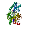
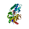
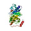
| Title | Structure of beta-phosphoglucomutase D10N mutant in complex with glucose-1,6-bisphosphate | ||||||
 Components Components | Beta-phosphoglucomutase | ||||||
 Keywords Keywords | ISOMERASE / phosphoryl transfer / catalysis / near attack complex | ||||||
| Function / homology |  Function and homology informationbeta-phosphoglucomutase / beta-phosphoglucomutase activity / carbohydrate metabolic process / magnesium ion binding / cytoplasm Function and homology informationbeta-phosphoglucomutase / beta-phosphoglucomutase activity / carbohydrate metabolic process / magnesium ion binding / cytoplasmSimilarity search - Function | ||||||
| Biological species |  Lactococcus lactis (lactic acid bacteria) Lactococcus lactis (lactic acid bacteria) | ||||||
| Method | X-RAY DIFFRACTION / SYNCHROTRON / MOLECULAR REPLACEMENT / Resolution: 2.2 Å | ||||||
 Authors Authors | Bowler, M.W. | ||||||
 Citation Citation | Journal: Acs Catalysis / Year: 2018 Title: van der Waals Contact between Nucleophile and Transferring Phosphorus Is Insufficient To Achieve Enzyme Transition-State Architecture Authors: Johnson, L.A. / Robertson, A.J. / Baxter, N.J. / Trevitt, C.R. / Bisson, C. / Jin, Y. / Wood, H.P. / Hounslow, A.M. / Cliff, M.J. / Blackburn, G.M. / Bowler, M.W. / Waltho, J.P. | ||||||
| History |
|
- Structure visualization
| Structure viewer | Molecule: MolmilJmol/JSmol |
|---|
- Downloads & links
-Download
| PDBx/mmCIF format | 5o6p.cif.gz | 99 KB | Display | PDBx/mmCIF format |
|---|---|---|---|---|
| PDB format | pdb5o6p.ent.gz | 74.5 KB | Display | PDB format |
| PDBx/mmJSON format | 5o6p.json.gz | Tree view | PDBx/mmJSON format | |
| Others |  Other downloads Other downloads |
-Validation report
| Arichive directory | https://data.pdbj.org/pub/pdb/validation_reports/o6/5o6pftp://data.pdbj.org/pub/pdb/validation_reports/o6/5o6p | HTTPS FTP |
|---|
-Related structure data
| Related structure data | 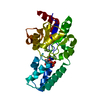 5o6rC  5ojzC  5ok0C 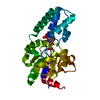 5ok1C 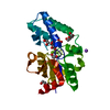 5ok2C 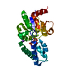 2wf9S S: Starting model for refinement C: citing same article ( |
|---|---|
| Similar structure data |


-Links
 PDBj
PDBj

- Assembly
| Deposited unit | 
| ||||||||
|---|---|---|---|---|---|---|---|---|---|
| 1 |
| ||||||||
| Unit cell |
|
-Components
| #1: Protein | / Beta-PGM Mass: 24238.609 Da / Num. of mol.: 1 / Mutation: D10N Source method: isolated from a genetically manipulated source Source: (gene. exp.) Lactococcus lactis (lactic acid bacteria)Strain: IL1403 / Gene: pgmB, LL0429, L0001 / Production host: Escherichia coli BL21(DE3) (bacteria) / References: UniProt: P71447, beta-phosphoglucomutase |
|---|---|
| #2: Sugar | ChemComp-B16 /   Type: D-saccharide / Mass: 340.116 Da / Num. of mol.: 1 Type: D-saccharide / Mass: 340.116 Da / Num. of mol.: 1Source method: isolated from a genetically manipulated source Formula: C6H14O12P2 / Feature type: SUBJECT OF INVESTIGATION |
| #3: Chemical | ChemComp-MG /   Mass: 24.305 Da / Num. of mol.: 1 / Source method: obtained synthetically / Formula: Mg Mass: 24.305 Da / Num. of mol.: 1 / Source method: obtained synthetically / Formula: Mg |
| #4: Water | ChemComp-HOH / Water Mass: 18.015 Da / Num. of mol.: 70 / Source method: isolated from a natural source / Formula: H2O Mass: 18.015 Da / Num. of mol.: 70 / Source method: isolated from a natural source / Formula: H2O |
-Experimental details
-Experiment
| Experiment | Method: X-RAY DIFFRACTION / Number of used crystals: 1 |
|---|
- Sample preparation
| Crystal | Density Matthews: 1.86 Å3/Da / Density % sol: 33.93 % Description: small needles with approximate dimensions 0.03 mm x 0.05 mm x 0.5 mm |
|---|---|
| Crystal grow | Temperature: 293 K / Method: vapor diffusion, sitting drop / pH: 7.2 / Details: 28-32 % PEG 4000 and 100 mM Na acetate |
-Data collection
| Diffraction | Mean temperature: 100 K |
|---|---|
| Diffraction source | Source: SYNCHROTRON / Site: ESRF  / Beamline: ID14-2 / Wavelength: 0.933 Å / Beamline: ID14-2 / Wavelength: 0.933 Å |
| Detector | Type: ADSC QUANTUM 210 / Detector: CCD / Date: Jan 30, 2009 / Details: toroidla mirror |
| Radiation | Monochromator: C001 / Protocol: SINGLE WAVELENGTH / Monochromatic (M) / Laue (L): M / Scattering type: x-ray |
| Radiation wavelength | Wavelength: 0.933 Å / Relative weight: 1 |
| Reflection | Resolution: 2.2→20 Å / Num. obs: 31189 / % possible obs: 98.2 % / Observed criterion σ(F): 3 / Observed criterion σ(I): 3 / Redundancy: 3.3 % / Biso Wilson estimate: 28.3 Å2 / Rmerge(I) obs: 0.08 / Net I/σ(I): 10 |
| Reflection shell | Resolution: 2.2→2.32 Å / Redundancy: 3.3 % / Rmerge(I) obs: 0.26 / Mean I/σ(I) obs: 4 / Num. unique obs: 4563 / Rpim(I) all: 0.17 / % possible all: 99.5 |
- Processing
| Software |
| ||||||||||||||||||||||||||||||||||||||||||||||||||||||||||||||||||||||||||||||||||||||||||||||||||||||||||||||||||||||||||||||||||||||||||||||||||||||||||||||||||||||||||||||||||||||
|---|---|---|---|---|---|---|---|---|---|---|---|---|---|---|---|---|---|---|---|---|---|---|---|---|---|---|---|---|---|---|---|---|---|---|---|---|---|---|---|---|---|---|---|---|---|---|---|---|---|---|---|---|---|---|---|---|---|---|---|---|---|---|---|---|---|---|---|---|---|---|---|---|---|---|---|---|---|---|---|---|---|---|---|---|---|---|---|---|---|---|---|---|---|---|---|---|---|---|---|---|---|---|---|---|---|---|---|---|---|---|---|---|---|---|---|---|---|---|---|---|---|---|---|---|---|---|---|---|---|---|---|---|---|---|---|---|---|---|---|---|---|---|---|---|---|---|---|---|---|---|---|---|---|---|---|---|---|---|---|---|---|---|---|---|---|---|---|---|---|---|---|---|---|---|---|---|---|---|---|---|---|---|---|
| Refinement | Method to determine structure: MOLECULAR REPLACEMENT Starting model: 2wf9 Resolution: 2.2→20 Å / Cor.coef. Fo:Fc: 0.943 / Cor.coef. Fo:Fc free: 0.912 / SU B: 15.032 / SU ML: 0.205 / Cross valid method: THROUGHOUT / ESU R: 0.391 / ESU R Free: 0.251 / Details: HYDROGENS HAVE BEEN USED IF PRESENT IN THE INPUT
| ||||||||||||||||||||||||||||||||||||||||||||||||||||||||||||||||||||||||||||||||||||||||||||||||||||||||||||||||||||||||||||||||||||||||||||||||||||||||||||||||||||||||||||||||||||||
| Solvent computation | Ion probe radii: 0.8 Å / Shrinkage radii: 0.8 Å / VDW probe radii: 1.2 Å | ||||||||||||||||||||||||||||||||||||||||||||||||||||||||||||||||||||||||||||||||||||||||||||||||||||||||||||||||||||||||||||||||||||||||||||||||||||||||||||||||||||||||||||||||||||||
| Displacement parameters | Biso mean: 39.635 Å2
| ||||||||||||||||||||||||||||||||||||||||||||||||||||||||||||||||||||||||||||||||||||||||||||||||||||||||||||||||||||||||||||||||||||||||||||||||||||||||||||||||||||||||||||||||||||||
| Refinement step | Cycle: 1 / Resolution: 2.2→20 Å
| ||||||||||||||||||||||||||||||||||||||||||||||||||||||||||||||||||||||||||||||||||||||||||||||||||||||||||||||||||||||||||||||||||||||||||||||||||||||||||||||||||||||||||||||||||||||
| Refine LS restraints |
|