presynaptic intermediate filament cytoskeleton / mitotic recombination-dependent replication fork processing / chromosome organization involved in meiotic cell cycle / cellular response to camptothecin / DNA recombinase assembly / telomere maintenance via telomere lengthening / positive regulation of DNA ligation / double-strand break repair involved in meiotic recombination / nuclear ubiquitin ligase complex / mitotic recombination ...presynaptic intermediate filament cytoskeleton / mitotic recombination-dependent replication fork processing / chromosome organization involved in meiotic cell cycle / cellular response to camptothecin / DNA recombinase assembly / telomere maintenance via telomere lengthening / positive regulation of DNA ligation / double-strand break repair involved in meiotic recombination / nuclear ubiquitin ligase complex / mitotic recombination / DNA strand invasion / cellular response to hydroxyurea / replication-born double-strand break repair via sister chromatid exchange / DNA strand exchange activity / lateral element / telomere maintenance via recombination / regulation of DNA damage checkpoint / Impaired BRCA2 binding to PALB2 / single-stranded DNA helicase activity / reciprocal meiotic recombination / Defective homologous recombination repair (HRR) due to BRCA1 loss of function / Defective HDR through Homologous Recombination Repair (HRR) due to PALB2 loss of BRCA1 binding function / Defective HDR through Homologous Recombination Repair (HRR) due to PALB2 loss of BRCA2/RAD51/RAD51C binding function / Homologous DNA Pairing and Strand Exchange / Resolution of D-loop Structures through Synthesis-Dependent Strand Annealing (SDSA) / Resolution of D-loop Structures through Holliday Junction Intermediates / HDR through Single Strand Annealing (SSA) / ATP-dependent DNA damage sensor activity / Impaired BRCA2 binding to RAD51 / regulation of double-strand break repair via homologous recombination / nuclear chromosome / DNA unwinding involved in DNA replication / replication fork processing / Transcriptional Regulation by E2F6 / Presynaptic phase of homologous DNA pairing and strand exchange / ATP-dependent activity, acting on DNA / interstrand cross-link repair / DNA polymerase binding / condensed chromosome / meiotic cell cycle / condensed nuclear chromosome / male germ cell nucleus / cellular response to ionizing radiation / double-strand break repair via homologous recombination / regulation of protein phosphorylation / HDR through Homologous Recombination (HRR) / PML body / Meiotic recombination / single-stranded DNA binding / site of double-strand break / double-stranded DNA binding / DNA recombination / chromosome, telomeric region / mitochondrial matrix / DNA repair / centrosome / DNA damage response / chromatin binding / chromatin / nucleolus / perinuclear region of cytoplasm / enzyme binding / ATP hydrolysis activity / protein-containing complex / mitochondrion / nucleoplasm / ATP binding / identical protein binding / nucleus / cytosol / cytoplasm Similarity search - Function
DNA recombination/repair protein Rad51 / DNA recombination and repair protein, RecA-like / DNA recombination and repair protein Rad51-like, C-terminal / Rad51 / DNA recombination and repair protein RecA, monomer-monomer interface / RecA family profile 2. / DNA recombination and repair protein RecA-like, ATP-binding domain / RecA family profile 1. / DNA repair Rad51/transcription factor NusA, alpha-helical / Helix-hairpin-helix domain ...DNA recombination/repair protein Rad51 / DNA recombination and repair protein, RecA-like / DNA recombination and repair protein Rad51-like, C-terminal / Rad51 / DNA recombination and repair protein RecA, monomer-monomer interface / RecA family profile 2. / DNA recombination and repair protein RecA-like, ATP-binding domain / RecA family profile 1. / DNA repair Rad51/transcription factor NusA, alpha-helical / Helix-hairpin-helix domain / 5' to 3' exonuclease, C-terminal subdomain / DNA polymerase; domain 1 / P-loop containing nucleotide triphosphate hydrolases / ATPases associated with a variety of cellular activities / AAA+ ATPase domain / P-loop containing nucleoside triphosphate hydrolase / Rossmann fold / Orthogonal Bundle / 3-Layer(aba) Sandwich / Mainly Alpha / Alpha Beta Similarity search - Domain/homology
#112 - Apr 2009 Oct and Sox Transcription Factors similarity (1)
-
Assembly
Deposited unit
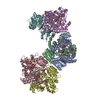
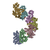
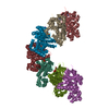
A: DNA repair protein RAD51 homolog 1 B: DNA repair protein RAD51 homolog 1 C: DNA repair protein RAD51 homolog 1 D: DNA repair protein RAD51 homolog 1 E: DNA repair protein RAD51 homolog 1 F: DNA repair protein RAD51 homolog 1 G: DNA repair protein RAD51 homolog 1 H: DNA repair protein RAD51 homolog 1 I: DNA repair protein RAD51 homolog 1 J: DNA repair protein RAD51 homolog 1 K: DNA repair protein RAD51 homolog 1 L: DNA repair protein RAD51 homolog 1 M: DNA repair protein RAD51 homolog 1 N: DNA repair protein RAD51 homolog 1 hetero molecules
A: DNA repair protein RAD51 homolog 1 B: DNA repair protein RAD51 homolog 1 C: DNA repair protein RAD51 homolog 1 D: DNA repair protein RAD51 homolog 1 E: DNA repair protein RAD51 homolog 1 F: DNA repair protein RAD51 homolog 1 G: DNA repair protein RAD51 homolog 1 hetero molecules
H: DNA repair protein RAD51 homolog 1 I: DNA repair protein RAD51 homolog 1 J: DNA repair protein RAD51 homolog 1 K: DNA repair protein RAD51 homolog 1 L: DNA repair protein RAD51 homolog 1 M: DNA repair protein RAD51 homolog 1 N: DNA repair protein RAD51 homolog 1 hetero molecules
In the structure databanks used in Yorodumi, some data are registered as the other names, "COVID-19 virus" and "2019-nCoV". Here are the details of the virus and the list of structure data.
Jan 31, 2019. EMDB accession codes are about to change! (news from PDBe EMDB page)
EMDB accession codes are about to change! (news from PDBe EMDB page)
The allocation of 4 digits for EMDB accession codes will soon come to an end. Whilst these codes will remain in use, new EMDB accession codes will include an additional digit and will expand incrementally as the available range of codes is exhausted. The current 4-digit format prefixed with “EMD-” (i.e. EMD-XXXX) will advance to a 5-digit format (i.e. EMD-XXXXX), and so on. It is currently estimated that the 4-digit codes will be depleted around Spring 2019, at which point the 5-digit format will come into force.
The EM Navigator/Yorodumi systems omit the EMD- prefix.
Related info.:Q: What is EMD? / ID/Accession-code notation in Yorodumi/EM Navigator
Yorodumi is a browser for structure data from EMDB, PDB, SASBDB, etc.
This page is also the successor to EM Navigator detail page, and also detail information page/front-end page for Omokage search.
The word "yorodu" (or yorozu) is an old Japanese word meaning "ten thousand". "mi" (miru) is to see.
Related info.:EMDB / PDB / SASBDB / Comparison of 3 databanks / Yorodumi Search / Aug 31, 2016. New EM Navigator & Yorodumi / Yorodumi Papers / Jmol/JSmol / Function and homology information / Changes in new EM Navigator and Yorodumi
 Movie
Movie Controller


 Open data
 Basic information
 Components
 Keywords
 Function and homology information
 Authors
 United Kingdom, 1items
  Citation
 Structure visualization
 Downloads & links
 Other downloads


 PDBj
PDBj


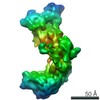
 Assembly


 Mass: 24.305 Da / Num. of mol.: 14
 Mass: 507.181 Da / Num. of mol.: 14
 Sample preparation
 / Beamline: PROXIMA 1 / Wavelength: 0.95373 Å
 Processing