 Movie
Movie Controller


+ Open data

- Basic information
| Entry | Database: PDB / ID: 5nw9 | ||||||
|---|---|---|---|---|---|---|---|
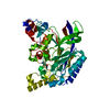
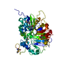
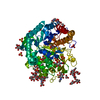
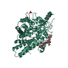
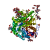
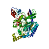
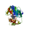
| Title | Crystal structure of the complex of Tdp1 with duplex DNA | ||||||
 Components Components |
| ||||||
 Keywords Keywords |  HYDROLASE / protein-DNA complex / DNA repair / Nucleosidase / phosphotyrosine diesterase HYDROLASE / protein-DNA complex / DNA repair / Nucleosidase / phosphotyrosine diesterase | ||||||
| Function / homology |  Function and homology information Function and homology information3'-tyrosyl-DNA phosphodiesterase activity / single strand break repair / Hydrolases; Acting on ester bonds; Phosphoric-diester hydrolases / exonuclease activity / Nonhomologous End-Joining (NHEJ) / double-strand break repair / single-stranded DNA binding / double-stranded DNA binding / intracellular membrane-bounded organelle / DNA repair ...3'-tyrosyl-DNA phosphodiesterase activity / single strand break repair / Hydrolases; Acting on ester bonds; Phosphoric-diester hydrolases / exonuclease activity / Nonhomologous End-Joining (NHEJ) / double-strand break repair / single-stranded DNA binding / double-stranded DNA binding / intracellular membrane-bounded organelle / DNA repair / nucleoplasm / nucleus / plasma membrane / cytoplasmSimilarity search - Function | ||||||
| Biological species |  Homo sapiens (human) Homo sapiens (human)synthetic construct (others) | ||||||
| Method | X-RAY DIFFRACTION / SYNCHROTRON / MOLECULAR REPLACEMENT / molecular replacement / Resolution: 2.04 Å | ||||||
| Model details | Tdp1 with product of nucleoside cleavage | ||||||
 Authors Authors | Richardson, J.M. / Ruksenaite, E. / Morris, E.R. | ||||||
| Funding support |  United Kingdom, 1items United Kingdom, 1items
| ||||||
 Citation Citation | Journal: Nat Commun / Year: 2018 Title: Structural basis for DNA 3'-end processing by human tyrosyl-DNA phosphodiesterase 1. Authors: Flett, F.J. / Ruksenaite, E. / Armstrong, L.A. / Bharati, S. / Carloni, R. / Morris, E.R. / Mackay, C.L. / Interthal, H. / Richardson, J.M. | ||||||
| History |
|
- Structure visualization
| Structure viewer | Molecule: MolmilJmol/JSmol |
|---|
- Downloads & links
-Download
| PDBx/mmCIF format | 5nw9.cif.gz | 363.3 KB | Display | PDBx/mmCIF format |
|---|---|---|---|---|
| PDB format | pdb5nw9.ent.gz | 293.3 KB | Display | PDB format |
| PDBx/mmJSON format | 5nw9.json.gz | Tree view | PDBx/mmJSON format | |
| Others |  Other downloads Other downloads |
-Validation report
| Arichive directory | https://data.pdbj.org/pub/pdb/validation_reports/nw/5nw9ftp://data.pdbj.org/pub/pdb/validation_reports/nw/5nw9 | HTTPS FTP |
|---|
-Related structure data
| Related structure data | 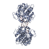 5nwaC  1jy1S S: Starting model for refinement C: citing same article ( |
|---|---|
| Similar structure data |


-Links
 PDBj
PDBj

- Assembly
| Deposited unit | 
| ||||||||
|---|---|---|---|---|---|---|---|---|---|
| 1 | 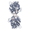
| ||||||||
| 2 | 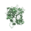
| ||||||||
| Unit cell |
|
-Components
| #1: Protein | Mass: 54731.195 Da / Num. of mol.: 2 / Fragment: delta 1-148 Source method: isolated from a genetically manipulated source Source: (gene. exp.) Homo sapiens (human) / Gene: TDP1 / Plasmid: pHN1894s D1-148 / Details (production host): pet15C / Production host:  Escherichia coli (E. coli) / Strain (production host): BL21(DE3) Escherichia coli (E. coli) / Strain (production host): BL21(DE3)References: UniProt: Q9NUW8, Hydrolases; Acting on ester bonds; Phosphoric-diester hydrolases#2: DNA chain | | Mass: 2755.823 Da / Num. of mol.: 1 / Source method: obtained synthetically / Details: IDT DNA synthesis / Source: (synth.) synthetic construct (others) #3: Water | ChemComp-HOH / | Water Mass: 18.015 Da / Num. of mol.: 57 / Source method: isolated from a natural source / Formula: H2O Mass: 18.015 Da / Num. of mol.: 57 / Source method: isolated from a natural source / Formula: H2O |
|---|
-Experimental details
-Experiment
| Experiment | Method: X-RAY DIFFRACTION / Number of used crystals: 1 |
|---|
- Sample preparation
| Crystal | Density Matthews: 2.39 Å3/Da / Density % sol: 48.51 % |
|---|---|
| Crystal grow | Temperature: 290 K / Method: vapor diffusion, sitting drop / pH: 7.5 Details: 0.1M Ammonium sulphate, 0.1M HEPES, 25% w/v PEG 8000 |
-Data collection
| Diffraction | Mean temperature: 100 K |
|---|---|
| Diffraction source | Source: SYNCHROTRON / Site: Diamond / Beamline: I04 / Wavelength: 0.9795 Å |
| Detector | Type: DECTRIS PILATUS3 S 6M / Detector: PIXEL / Date: Apr 5, 2015 |
| Radiation | Protocol: SINGLE WAVELENGTH / Monochromatic (M) / Laue (L): M / Scattering type: x-ray |
| Radiation wavelength | Wavelength: 0.9795 Å / Relative weight: 1 |
| Reflection | Resolution: 2.04→52.05 Å / Num. obs: 69045 / % possible obs: 99.2 % / Redundancy: 9.6 % / Net I/σ(I): 15.3 |
-Phasing
| Phasing | Method: molecular replacement |
|---|
- Processing
| Software |
| ||||||||||||||||||||||||||||||||||||||||||||||||||||||||||||||||||||||||||||||||||||||||||||||||||||
|---|---|---|---|---|---|---|---|---|---|---|---|---|---|---|---|---|---|---|---|---|---|---|---|---|---|---|---|---|---|---|---|---|---|---|---|---|---|---|---|---|---|---|---|---|---|---|---|---|---|---|---|---|---|---|---|---|---|---|---|---|---|---|---|---|---|---|---|---|---|---|---|---|---|---|---|---|---|---|---|---|---|---|---|---|---|---|---|---|---|---|---|---|---|---|---|---|---|---|---|---|---|
| Refinement | Method to determine structure: MOLECULAR REPLACEMENT Starting model: pdbid 1JY1 Resolution: 2.04→52.05 Å / Cor.coef. Fo:Fc: 0.954 / Cor.coef. Fo:Fc free: 0.943 / SU B: 17.861 / SU ML: 0.194 / Cross valid method: THROUGHOUT / σ(F): 0 / ESU R: 0.211 / ESU R Free: 0.186 / Details: molecular replacement
| ||||||||||||||||||||||||||||||||||||||||||||||||||||||||||||||||||||||||||||||||||||||||||||||||||||
| Solvent computation | Ion probe radii: 0.8 Å / Shrinkage radii: 0.8 Å / VDW probe radii: 1.2 Å | ||||||||||||||||||||||||||||||||||||||||||||||||||||||||||||||||||||||||||||||||||||||||||||||||||||
| Displacement parameters | Biso max: 144.24 Å2 / Biso mean: 56.481 Å2 / Biso min: 29.32 Å2
| ||||||||||||||||||||||||||||||||||||||||||||||||||||||||||||||||||||||||||||||||||||||||||||||||||||
| Refinement step | Cycle: final / Resolution: 2.04→52.05 Å
| ||||||||||||||||||||||||||||||||||||||||||||||||||||||||||||||||||||||||||||||||||||||||||||||||||||
| Refine LS restraints |
| ||||||||||||||||||||||||||||||||||||||||||||||||||||||||||||||||||||||||||||||||||||||||||||||||||||
| LS refinement shell | Resolution: 2.04→2.093 Å / Rfactor Rfree error: 0 / Total num. of bins used: 20
| ||||||||||||||||||||||||||||||||||||||||||||||||||||||||||||||||||||||||||||||||||||||||||||||||||||
| Refinement TLS params. | Method: refined / Refine-ID: X-RAY DIFFRACTION
| ||||||||||||||||||||||||||||||||||||||||||||||||||||||||||||||||||||||||||||||||||||||||||||||||||||
| Refinement TLS group |
|
