 Movie
Movie Controller


+ Open data

- Basic information
| Entry | Database: PDB / ID: 5nvl | ||||||
|---|---|---|---|---|---|---|---|
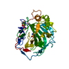
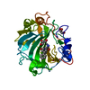
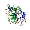
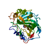
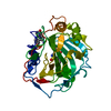
| Title | Crystal structure of the human 4EHP-GIGYF2 complex | ||||||
 Components Components |
| ||||||
 Keywords Keywords |  TRANSLATION / translational regulation / cap-binding protein / 4EHP-binding protein / GRB10-interacting GYF protein 2 TRANSLATION / translational regulation / cap-binding protein / 4EHP-binding protein / GRB10-interacting GYF protein 2 | ||||||
| Function / homology |  Function and homology information Function and homology informationmusculoskeletal movement / post-transcriptional gene silencing / spinal cord motor neuron differentiation / proximal dendrite / RNA cap binding / eukaryotic translation initiation factor 4F complex / translation factor activity, RNA binding / mRNA cap binding / miRNA-mediated gene silencing by inhibition of translation / RNA 7-methylguanosine cap binding ...musculoskeletal movement / post-transcriptional gene silencing / spinal cord motor neuron differentiation / proximal dendrite / RNA cap binding / eukaryotic translation initiation factor 4F complex / translation factor activity, RNA binding / mRNA cap binding / miRNA-mediated gene silencing by inhibition of translation / RNA 7-methylguanosine cap binding / feeding behavior / proline-rich region binding / negative regulation of type I interferon-mediated signaling pathway / mRNA destabilization / neuromuscular process controlling balance / negative regulation of translational initiation / homeostasis of number of cells within a tissue / translational initiation / mitotic G1 DNA damage checkpoint signaling / translation initiation factor activity / rescue of stalled ribosome / adult locomotory behavior / insulin-like growth factor receptor signaling pathway / post-embryonic development / P-body / multicellular organism growth / ISG15 antiviral mechanism / cytoplasmic stress granule / perikaryon / vesicle / molecular adaptor activity / negative regulation of translation / endosome / cadherin binding / ubiquitin protein ligase binding / Golgi apparatus / endoplasmic reticulum / protein-containing complex / RNA binding / membrane / cytosol / cytoplasmSimilarity search - Function | ||||||
| Biological species |  Homo sapiens (human) Homo sapiens (human) | ||||||
| Method | X-RAY DIFFRACTION / SYNCHROTRON / MOLECULAR REPLACEMENT / Resolution: 2.3 Å | ||||||
 Authors Authors | Peter, D. / Valkov, E. | ||||||
 Citation Citation | Journal: Genes Dev. / Year: 2017 Title: GIGYF1/2 proteins use auxiliary sequences to selectively bind to 4EHP and repress target mRNA expression. Authors: Peter, D. / Weber, R. / Sandmeir, F. / Wohlbold, L. / Helms, S. / Bawankar, P. / Valkov, E. / Igreja, C. / Izaurralde, E. | ||||||
| History |
|
- Structure visualization
| Structure viewer | Molecule: MolmilJmol/JSmol |
|---|
- Downloads & links
-Download
| PDBx/mmCIF format | 5nvl.cif.gz | 279.7 KB | Display | PDBx/mmCIF format |
|---|---|---|---|---|
| PDB format | pdb5nvl.ent.gz | 234.1 KB | Display | PDB format |
| PDBx/mmJSON format | 5nvl.json.gz | Tree view | PDBx/mmJSON format | |
| Others |  Other downloads Other downloads |
-Validation report
| Arichive directory | https://data.pdbj.org/pub/pdb/validation_reports/nv/5nvlftp://data.pdbj.org/pub/pdb/validation_reports/nv/5nvl | HTTPS FTP |
|---|
-Related structure data
| Related structure data | 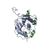 5nvkC 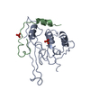 5nvmC  5nvnC 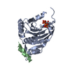 2jgbS S: Starting model for refinement C: citing same article ( |
|---|---|
| Similar structure data |


-Links
 PDBj
PDBj
- Assembly
| Deposited unit | 
| ||||||||
|---|---|---|---|---|---|---|---|---|---|
| 1 | 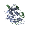
| ||||||||
| 2 | 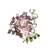
| ||||||||
| Unit cell |
|
-Components
| #1: Protein | Mass: 21979.102 Da / Num. of mol.: 2 Source method: isolated from a genetically manipulated source Details: the first 5 residues of the coordinate sequence of chain A and the first residue of the coordinate sequence of chain C belong to the expression tag Source: (gene. exp.) Homo sapiens (human) / Gene: EIF4E2, EIF4EL3 / Production host:  Escherichia coli BL21(DE3) (bacteria) / Variant (production host): STAR / References: UniProt: O60573 Escherichia coli BL21(DE3) (bacteria) / Variant (production host): STAR / References: UniProt: O60573#2: Protein | Mass: 8689.040 Da / Num. of mol.: 2 Source method: isolated from a genetically manipulated source Source: (gene. exp.) Homo sapiens (human) / Gene: GIGYF2, KIAA0642, PERQ2, TNRC15 / Production host: Escherichia coli BL21(DE3) (bacteria) / Variant (production host): STAR / References: UniProt: Q6Y7W6#3: Water | ChemComp-HOH / | Water Mass: 18.015 Da / Num. of mol.: 70 / Source method: isolated from a natural source / Formula: H2O Mass: 18.015 Da / Num. of mol.: 70 / Source method: isolated from a natural source / Formula: H2O |
|---|
-Experimental details
-Experiment
| Experiment | Method: X-RAY DIFFRACTION / Number of used crystals: 1 |
|---|
- Sample preparation
| Crystal | Density Matthews: 2.06 Å3/Da / Density % sol: 40.35 % |
|---|---|
| Crystal grow | Temperature: 291 K / Method: vapor diffusion, hanging drop Details: 0.1 M sodium citrate pH 5.0 0.1 M magnesium chloride 12% PEG 4000 |
-Data collection
| Diffraction | Mean temperature: 100 K |
|---|---|
| Diffraction source | Source: SYNCHROTRON / Site: SLS  / Beamline: X10SA / Wavelength: 1.00001 Å / Beamline: X10SA / Wavelength: 1.00001 Å |
| Detector | Type: PSI PILATUS 6M / Detector: PIXEL / Date: Feb 19, 2015 |
| Radiation | Protocol: SINGLE WAVELENGTH / Monochromatic (M) / Laue (L): M / Scattering type: x-ray |
| Radiation wavelength | Wavelength: 1.00001 Å / Relative weight: 1 |
| Reflection | Resolution: 2.3→45.9 Å / Num. obs: 23500 / % possible obs: 99.8 % / Redundancy: 11.2 % / Rsym value: 0.125 / Net I/σ(I): 13.2 |
| Reflection shell | Resolution: 2.3→2.36 Å / Redundancy: 10.6 % / Mean I/σ(I) obs: 2.06 / Num. unique all: 1690 / Rsym value: 0.938 / % possible all: 98.3 |
- Processing
| Software |
| |||||||||||||||||||||||||||||||||||||||||||||||||||||||||||||||||||||||||||||||||||||||||||||||||||||||||||||||||||||||||||||
|---|---|---|---|---|---|---|---|---|---|---|---|---|---|---|---|---|---|---|---|---|---|---|---|---|---|---|---|---|---|---|---|---|---|---|---|---|---|---|---|---|---|---|---|---|---|---|---|---|---|---|---|---|---|---|---|---|---|---|---|---|---|---|---|---|---|---|---|---|---|---|---|---|---|---|---|---|---|---|---|---|---|---|---|---|---|---|---|---|---|---|---|---|---|---|---|---|---|---|---|---|---|---|---|---|---|---|---|---|---|---|---|---|---|---|---|---|---|---|---|---|---|---|---|---|---|---|
| Refinement | Method to determine structure: MOLECULAR REPLACEMENT Starting model: 2JGB Resolution: 2.3→45.888 Å / SU ML: 0.32 / Cross valid method: FREE R-VALUE / σ(F): 1.37 / Phase error: 27.9
| |||||||||||||||||||||||||||||||||||||||||||||||||||||||||||||||||||||||||||||||||||||||||||||||||||||||||||||||||||||||||||||
| Solvent computation | Shrinkage radii: 0.9 Å / VDW probe radii: 1.11 Å | |||||||||||||||||||||||||||||||||||||||||||||||||||||||||||||||||||||||||||||||||||||||||||||||||||||||||||||||||||||||||||||
| Displacement parameters | Biso mean: 62 Å2 | |||||||||||||||||||||||||||||||||||||||||||||||||||||||||||||||||||||||||||||||||||||||||||||||||||||||||||||||||||||||||||||
| Refinement step | Cycle: LAST / Resolution: 2.3→45.888 Å
| |||||||||||||||||||||||||||||||||||||||||||||||||||||||||||||||||||||||||||||||||||||||||||||||||||||||||||||||||||||||||||||
| Refine LS restraints |
| |||||||||||||||||||||||||||||||||||||||||||||||||||||||||||||||||||||||||||||||||||||||||||||||||||||||||||||||||||||||||||||
| LS refinement shell |
| |||||||||||||||||||||||||||||||||||||||||||||||||||||||||||||||||||||||||||||||||||||||||||||||||||||||||||||||||||||||||||||
| Refinement TLS params. | Method: refined / Refine-ID: X-RAY DIFFRACTION
| |||||||||||||||||||||||||||||||||||||||||||||||||||||||||||||||||||||||||||||||||||||||||||||||||||||||||||||||||||||||||||||
| Refinement TLS group |
|
