- PDB-5nvd: Crystal structure of hexameric CBS-CP12 protein from bloom-formin... -
+
Open data
ID or keywords:
Loading...
-
Basic information
Entry
Database: PDB / ID: 5nvd
Title
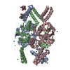
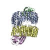
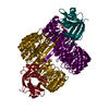
Crystal structure of hexameric CBS-CP12 protein from bloom-forming cyanobacteria at 2.5 A resolution in P6322 crystal form
Components
CBS-CP12
Keywords
PHOTOSYNTHESIS / Cystathionine beta synthase domain / fusion protein / redox-regulation of photosynthesis
Function / homology
CP12 domain / CP12 domain / CP12 / CBS domain superfamily / Domain in cystathionine beta-synthase and other proteins. / CBS domain / CBS domain / CBS domain profile. / Similar to tr
Mass: 23154.561 Da / Num. of mol.: 1 Source method: isolated from a genetically manipulated source Details: First four residues in the sequence are EXPRESSION TAG Source: (gene. exp.) Microcystis aeruginosa PCC 7806 (bacteria) Gene: IPF_2164 / Production host: Escherichia coli (E. coli) / Strain (production host): LOBSTR / References: UniProt: A8YJ50
Mass: 18.015 Da / Num. of mol.: 31 / Source method: isolated from a natural source / Formula: H2O
-
Experimental details
-
Experiment
Experiment
Method: X-RAY DIFFRACTION / Number of used crystals: 1
-
Sample preparation
Crystal
Density Matthews: 3.03 Å3/Da / Density % sol: 60 %
Crystal grow
Temperature: 293 K / Method: vapor diffusion, hanging drop / pH: 7 Details: Protein at 6.08 mg/ml in 50 mM Bicine/KOH pH 7.8, 40 mM KCl, 0.132 mM AMP solution was mixed with reservoir solution containing 0.15 M KSCN, 0.1 M HEPES pH 7.0 and 18% PEG3350 containing 0.1 ...Details: Protein at 6.08 mg/ml in 50 mM Bicine/KOH pH 7.8, 40 mM KCl, 0.132 mM AMP solution was mixed with reservoir solution containing 0.15 M KSCN, 0.1 M HEPES pH 7.0 and 18% PEG3350 containing 0.1 mM Guanidine hydrochloride. Cryoprotector - reservoir solution with 25% ethylene glycol.
-
Data collection
Diffraction
Mean temperature: 100 K
Diffraction source
Source: SYNCHROTRON / Site: PETRA III, EMBL c/o DESY / Beamline: P13 (MX1) / Wavelength: 0.9184 Å
Resolution: 2.5→85.07 Å / Cor.coef. Fo:Fc: 0.949 / Cor.coef. Fo:Fc free: 0.937 / SU B: 20.157 / SU ML: 0.205 / Cross valid method: THROUGHOUT / ESU R: 0.284 / ESU R Free: 0.223 / Details: HYDROGENS HAVE BEEN ADDED IN THE RIDING POSITIONS
Rfactor
Num. reflection
% reflection
Selection details
Rfree
0.23446
495
4.8 %
RANDOM
Rwork
0.19866
-
-
-
obs
0.20038
9924
99.94 %
-
Solvent computation
Ion probe radii: 0.8 Å / Shrinkage radii: 0.8 Å / VDW probe radii: 1.2 Å
 Movie
Movie Controller

 Yorodumi
 Open data
 Basic information
 Components
 Keywords
 PHOTOSYNTHESIS /
  Function and homology information
 Authors
 Citation
 Structure visualization
 Downloads & links
 Other downloads
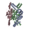


 PDBj
PDBj
 Assembly
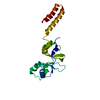

 Mass: 18.015 Da / Num. of mol.: 31 / Source method: isolated from a natural source / Formula: H2O
 Sample preparation
 / Beamline: P13 (MX1) / Wavelength: 0.9184 Å
 Processing