 Movie
Movie Controller


+ Open data

- Basic information
| Entry | Database: PDB / ID: 5nty | ||||||
|---|---|---|---|---|---|---|---|
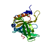
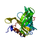
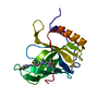
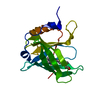
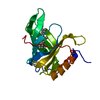
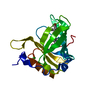
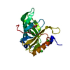
| Title | Structure of non-fluorescent human plasma RBP4 | ||||||
 Components Components | Retinol-binding protein 4 | ||||||
 Keywords Keywords |  TRANSPORT PROTEIN / RBP4 / plasma retinol-binding protein / lipocalin / fatty acid / signaling protein TRANSPORT PROTEIN / RBP4 / plasma retinol-binding protein / lipocalin / fatty acid / signaling protein | ||||||
| Function / homology |  Function and homology information Function and homology informationRetinoid metabolism disease events / urinary bladder development / embryonic retina morphogenesis in camera-type eye / retinol transport / female genitalia morphogenesis / retinol transmembrane transporter activity / embryonic organ morphogenesis / maintenance of gastrointestinal epithelium / embryonic skeletal system development / negative regulation of cardiac muscle cell proliferation ...Retinoid metabolism disease events / urinary bladder development / embryonic retina morphogenesis in camera-type eye / retinol transport / female genitalia morphogenesis / retinol transmembrane transporter activity / embryonic organ morphogenesis / maintenance of gastrointestinal epithelium / embryonic skeletal system development / negative regulation of cardiac muscle cell proliferation / eye development / heart trabecula formation / cardiac muscle tissue development / retinal binding / retinol metabolic process / retinol binding / positive regulation of immunoglobulin production / Retinoid cycle disease events / The canonical retinoid cycle in rods (twilight vision) / uterus development / vagina development / response to retinoic acid / Retinoid metabolism and transport / visual perception / gluconeogenesis / lung development / positive regulation of insulin secretion / glucose homeostasis / heart development / extracellular space / extracellular exosome / extracellular regionSimilarity search - Function | ||||||
| Biological species |  Homo sapiens (human) Homo sapiens (human) | ||||||
| Method | X-RAY DIFFRACTION / SYNCHROTRON / MOLECULAR REPLACEMENT / Resolution: 2 Å | ||||||
 Authors Authors | Perduca, M. / Monaco, H.L. / Galliano, M. | ||||||
 Citation Citation | Journal: Biochim. Biophys. Acta / Year: 2018 Title: Human plasma retinol-binding protein (RBP4) is also a fatty acid-binding protein. Authors: Perduca, M. / Nicolis, S. / Mannucci, B. / Galliano, M. / Monaco, H.L. | ||||||
| History |
|
- Structure visualization
| Structure viewer | Molecule: MolmilJmol/JSmol |
|---|
- Downloads & links
-Download
| PDBx/mmCIF format | 5nty.cif.gz | 90.9 KB | Display | PDBx/mmCIF format |
|---|---|---|---|---|
| PDB format | pdb5nty.ent.gz | 68.4 KB | Display | PDB format |
| PDBx/mmJSON format | 5nty.json.gz | Tree view | PDBx/mmJSON format | |
| Others |  Other downloads Other downloads |
-Validation report
| Arichive directory | https://data.pdbj.org/pub/pdb/validation_reports/nt/5ntyftp://data.pdbj.org/pub/pdb/validation_reports/nt/5nty | HTTPS FTP |
|---|
-Related structure data
| Related structure data | 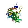 5nu2C 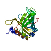 5nu6C  5nu7C  5nu8C 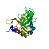 5nu9C 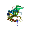 5nuaC  5nubC  1brqS S: Starting model for refinement C: citing same article ( |
|---|---|
| Similar structure data |


-Links
 PDBj
PDBj


- Assembly
| Deposited unit | 
| ||||||||
|---|---|---|---|---|---|---|---|---|---|
| 1 |
| ||||||||
| Unit cell |
|
-Components
| #1: Protein | Mass: 20984.445 Da / Num. of mol.: 1 / Source method: isolated from a natural source / Source: (natural) Homo sapiens (human) / References: UniProt: P02753 |
|---|---|
| #2: Chemical | ChemComp-PLM / Palmitic acid  Mass: 256.424 Da / Num. of mol.: 1 / Source method: obtained synthetically / Formula: C16H32O2 Mass: 256.424 Da / Num. of mol.: 1 / Source method: obtained synthetically / Formula: C16H32O2 |
| #3: Chemical | ChemComp-CL / Chloride  Mass: 35.453 Da / Num. of mol.: 1 / Source method: obtained synthetically / Formula: Cl Mass: 35.453 Da / Num. of mol.: 1 / Source method: obtained synthetically / Formula: Cl |
| #4: Water | ChemComp-HOH / Water Mass: 18.015 Da / Num. of mol.: 78 / Source method: isolated from a natural source / Formula: H2O Mass: 18.015 Da / Num. of mol.: 78 / Source method: isolated from a natural source / Formula: H2O |
-Experimental details
-Experiment
| Experiment | Method: X-RAY DIFFRACTION / Number of used crystals: 1 |
|---|
- Sample preparation
| Crystal | Density Matthews: 3.65 Å3/Da / Density % sol: 66.31 % |
|---|---|
| Crystal grow | Temperature: 279 K / Method: microdialysis / Details: 4.5M NaCl, 20mM Na cacodylate, pH 6.8 |
-Data collection
| Diffraction | Mean temperature: 100 K |
|---|---|
| Diffraction source | Source: SYNCHROTRON / Site: ESRF  / Beamline: ID29 / Wavelength: 1 Å / Beamline: ID29 / Wavelength: 1 Å |
| Detector | Type: DECTRIS PILATUS 6M-F / Detector: PIXEL / Date: Jul 16, 2015 |
| Radiation | Protocol: SINGLE WAVELENGTH / Monochromatic (M) / Laue (L): M / Scattering type: x-ray |
| Radiation wavelength | Wavelength: 1 Å / Relative weight: 1 |
| Reflection | Resolution: 2→40 Å / Num. obs: 19780 / % possible obs: 98.5 % / Redundancy: 4.4 % / Rmerge(I) obs: 0.106 / Net I/σ(I): 7.9 |
| Reflection shell | Resolution: 2→2.11 Å / Redundancy: 4.2 % / Rmerge(I) obs: 0.491 / Mean I/σ(I) obs: 2.5 / % possible all: 98.3 |
- Processing
| Software |
| ||||||||||||||||||||||||||||||||||||||||||||||||||||||||||||||||||||||||||||||||||||||||||||||||||||||||||||||||||||||||||||||||||||||||||||||||||||||||||||||||||||||||||||||||||||||
|---|---|---|---|---|---|---|---|---|---|---|---|---|---|---|---|---|---|---|---|---|---|---|---|---|---|---|---|---|---|---|---|---|---|---|---|---|---|---|---|---|---|---|---|---|---|---|---|---|---|---|---|---|---|---|---|---|---|---|---|---|---|---|---|---|---|---|---|---|---|---|---|---|---|---|---|---|---|---|---|---|---|---|---|---|---|---|---|---|---|---|---|---|---|---|---|---|---|---|---|---|---|---|---|---|---|---|---|---|---|---|---|---|---|---|---|---|---|---|---|---|---|---|---|---|---|---|---|---|---|---|---|---|---|---|---|---|---|---|---|---|---|---|---|---|---|---|---|---|---|---|---|---|---|---|---|---|---|---|---|---|---|---|---|---|---|---|---|---|---|---|---|---|---|---|---|---|---|---|---|---|---|---|---|
| Refinement | Method to determine structure: MOLECULAR REPLACEMENT Starting model: 1brq Resolution: 2→30 Å / Cor.coef. Fo:Fc: 0.967 / Cor.coef. Fo:Fc free: 0.939 / SU B: 10.31 / SU ML: 0.122 / Cross valid method: THROUGHOUT / ESU R: 0.188 / ESU R Free: 0.133
| ||||||||||||||||||||||||||||||||||||||||||||||||||||||||||||||||||||||||||||||||||||||||||||||||||||||||||||||||||||||||||||||||||||||||||||||||||||||||||||||||||||||||||||||||||||||
| Solvent computation | Ion probe radii: 0.8 Å / Shrinkage radii: 0.8 Å / VDW probe radii: 1.2 Å | ||||||||||||||||||||||||||||||||||||||||||||||||||||||||||||||||||||||||||||||||||||||||||||||||||||||||||||||||||||||||||||||||||||||||||||||||||||||||||||||||||||||||||||||||||||||
| Displacement parameters | Biso mean: 55.543 Å2
| ||||||||||||||||||||||||||||||||||||||||||||||||||||||||||||||||||||||||||||||||||||||||||||||||||||||||||||||||||||||||||||||||||||||||||||||||||||||||||||||||||||||||||||||||||||||
| Refinement step | Cycle: 1 / Resolution: 2→30 Å
| ||||||||||||||||||||||||||||||||||||||||||||||||||||||||||||||||||||||||||||||||||||||||||||||||||||||||||||||||||||||||||||||||||||||||||||||||||||||||||||||||||||||||||||||||||||||
| Refine LS restraints |
|