 Movie
Movie Controller

[English] 日本語
 Yorodumi
- PDB-5nsq: Structure of Leucyl aminopeptidase from Trypanosoma brucei in com... -
+ Open data

- Basic information
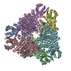
| Entry | Database: PDB / ID: 5nsq | ||||||
|---|---|---|---|---|---|---|---|
| Title | Structure of Leucyl aminopeptidase from Trypanosoma brucei in complex with Actinonin | ||||||
 Components Components | Aminopeptidase, putative | ||||||
 Keywords Keywords | HYDROLASE / M17 leucyl aminopeptidase / aminopeptidase / tryanosoma brucei actinonin | ||||||
| Function / homology |  Function and homology information Function and homology informationmetalloaminopeptidase activity / aminopeptidase activity / manganese ion binding / peptidase activity / proteolysis / cytoplasmSimilarity search - Function | ||||||
| Biological species |  Trypanosoma brucei brucei TREU927 (eukaryote) Trypanosoma brucei brucei TREU927 (eukaryote) | ||||||
| Method | X-RAY DIFFRACTION / SYNCHROTRON / MOLECULAR REPLACEMENT / Resolution: 3 Å | ||||||
 Authors Authors | Timm, J. / Wilson, K. | ||||||
 Citation Citation | Journal: mSphere / Year: 2018 Title: Structural Characterization of Acidic M17 Leucine Aminopeptidases from the TriTryps and Evaluation of Their Role in Nutrient Starvation inTrypanosoma brucei. Authors: Timm, J. / Valente, M. / Garcia-Caballero, D. / Wilson, K.S. / Gonzalez-Pacanowska, D. | ||||||
| History |
|
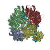
- Structure visualization
| Structure viewer | Molecule: MolmilJmol/JSmol |
|---|
- Downloads & links
-Download
| PDBx/mmCIF format | 5nsq.cif.gz | 559.6 KB | Display | PDBx/mmCIF format |
|---|---|---|---|---|
| PDB format | pdb5nsq.ent.gz | 459.5 KB | Display | PDB format |
| PDBx/mmJSON format | 5nsq.json.gz | Tree view | PDBx/mmJSON format | |
| Others |  Other downloads Other downloads |
-Validation report
| Arichive directory | https://data.pdbj.org/pub/pdb/validation_reports/ns/5nsqftp://data.pdbj.org/pub/pdb/validation_reports/ns/5nsq | HTTPS FTP |
|---|
-Related structure data
| Related structure data | 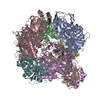 5nskSC  5nsmC 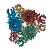 5ntdC  5ntfC  5ntgC 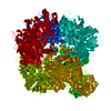 5nthC S: Starting model for refinement C: citing same article ( |
|---|---|
| Similar structure data |


-Links
 PDBj
PDBj- Assembly
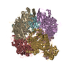
| Deposited unit | 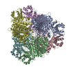
| |||||||||||||||||||||||||||||||||||||||||||||||||||||||||||||||||||||||||||||||||||||||||||||||||||||||||||||||||||||||||||||||||||||||||||||||||||||||||||||||||||||||||||||||||||||||||||||||||||||||||||||||||||||||||||||||||||||||
|---|---|---|---|---|---|---|---|---|---|---|---|---|---|---|---|---|---|---|---|---|---|---|---|---|---|---|---|---|---|---|---|---|---|---|---|---|---|---|---|---|---|---|---|---|---|---|---|---|---|---|---|---|---|---|---|---|---|---|---|---|---|---|---|---|---|---|---|---|---|---|---|---|---|---|---|---|---|---|---|---|---|---|---|---|---|---|---|---|---|---|---|---|---|---|---|---|---|---|---|---|---|---|---|---|---|---|---|---|---|---|---|---|---|---|---|---|---|---|---|---|---|---|---|---|---|---|---|---|---|---|---|---|---|---|---|---|---|---|---|---|---|---|---|---|---|---|---|---|---|---|---|---|---|---|---|---|---|---|---|---|---|---|---|---|---|---|---|---|---|---|---|---|---|---|---|---|---|---|---|---|---|---|---|---|---|---|---|---|---|---|---|---|---|---|---|---|---|---|---|---|---|---|---|---|---|---|---|---|---|---|---|---|---|---|---|---|---|---|---|---|---|---|---|---|---|---|---|---|---|---|---|---|
| 1 |
| |||||||||||||||||||||||||||||||||||||||||||||||||||||||||||||||||||||||||||||||||||||||||||||||||||||||||||||||||||||||||||||||||||||||||||||||||||||||||||||||||||||||||||||||||||||||||||||||||||||||||||||||||||||||||||||||||||||||
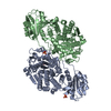
| Unit cell |
| |||||||||||||||||||||||||||||||||||||||||||||||||||||||||||||||||||||||||||||||||||||||||||||||||||||||||||||||||||||||||||||||||||||||||||||||||||||||||||||||||||||||||||||||||||||||||||||||||||||||||||||||||||||||||||||||||||||||
| Noncrystallographic symmetry (NCS) | NCS domain:
NCS domain segments: Component-ID: 0 / Refine code: 0
|