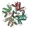
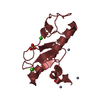
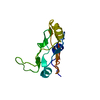
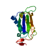
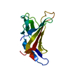
Journal: Acta Crystallogr F Struct Biol Commun / Year: 2018 Title: Structure and stability of the Human respiratory syncytial virus M RNA-binding core domain reveals a compact and cooperative folding unit. Authors: Ivana G Molina / Inokentijs Josts / Yasser Almeida Hernandez / Sebastian Esperante / Mariano Salgueiro / Maria M Garcia Alai / Gonzalo de Prat-Gay / Henning Tidow / Abstract: Human syncytial respiratory virus is a nonsegmented negative-strand RNA virus with serious implications for respiratory disease in infants, and has recently been reclassified into a new family, ...Human syncytial respiratory virus is a nonsegmented negative-strand RNA virus with serious implications for respiratory disease in infants, and has recently been reclassified into a new family, Pneumoviridae. One of the main reasons for this classification is the unique presence of a transcriptional antiterminator, called M. The puzzling mechanism of action of M, which is a rarity among antiterminators in viruses and is part of the RNA polymerase complex, relies on dissecting the structure and function of this multidomain tetramer. The RNA-binding activity is located in a monomeric globular `core' domain, a high-resolution crystal structure of which is now presented. The structure reveals a compact domain which is superimposable on the full-length M tetramer, with additional electron density for the C-terminal tail that was not observed in the previous models. Moreover, its folding stability was determined through chemical denaturation, which shows that the secondary and tertiary structure unfold concomitantly, which is indicative of a two-state equilibrium. These results constitute a further step in the understanding of this unique RNA-binding domain, for which there is no sequence or structural counterpart outside this virus family, in addition to its implications in transcription regulation and its likeliness as an antiviral target.
In the structure databanks used in Yorodumi, some data are registered as the other names, "COVID-19 virus" and "2019-nCoV". Here are the details of the virus and the list of structure data.
Jan 31, 2019. EMDB accession codes are about to change! (news from PDBe EMDB page)
EMDB accession codes are about to change! (news from PDBe EMDB page)
The allocation of 4 digits for EMDB accession codes will soon come to an end. Whilst these codes will remain in use, new EMDB accession codes will include an additional digit and will expand incrementally as the available range of codes is exhausted. The current 4-digit format prefixed with “EMD-” (i.e. EMD-XXXX) will advance to a 5-digit format (i.e. EMD-XXXXX), and so on. It is currently estimated that the 4-digit codes will be depleted around Spring 2019, at which point the 5-digit format will come into force.
The EM Navigator/Yorodumi systems omit the EMD- prefix.
Related info.:Q: What is EMD? / ID/Accession-code notation in Yorodumi/EM Navigator
Yorodumi is a browser for structure data from EMDB, PDB, SASBDB, etc.
This page is also the successor to EM Navigator detail page, and also detail information page/front-end page for Omokage search.
The word "yorodu" (or yorozu) is an old Japanese word meaning "ten thousand". "mi" (miru) is to see.
Related info.:EMDB / PDB / SASBDB / Comparison of 3 databanks / Yorodumi Search / Aug 31, 2016. New EM Navigator & Yorodumi / Yorodumi Papers / Jmol/JSmol / Function and homology information / Changes in new EM Navigator and Yorodumi
 Movie
Movie Controller


 Open data
 Basic information
 Components
 Keywords
 VIRAL PROTEIN
 Function and homology information
 Authors
 Citation

 Structure visualization
 Downloads & links
 Other downloads
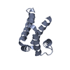


 PDBj
PDBj
 Assembly


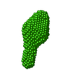
 Mass: 18.015 Da / Num. of mol.: 189 / Source method: isolated from a natural source / Formula: H2O
 Sample preparation
 Processing