 Movie
Movie Controller

[English] 日本語
 Yorodumi
- PDB-5ncv: Crystal Structure of Cytochrome c in complex with p-Methylphospho... -
+ Open data

- Basic information
| Entry | Database: PDB / ID: 5ncv | ||||||
|---|---|---|---|---|---|---|---|
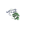
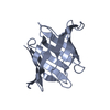
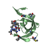
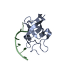
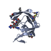
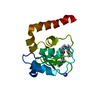
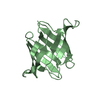
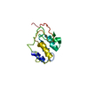
| Title | Crystal Structure of Cytochrome c in complex with p-Methylphosphonatocalix[4]arene | ||||||
 Components Components | Cytochrome c iso-1 | ||||||
 Keywords Keywords |  OXIDOREDUCTASE / methylphosphonatocalix[4]arene / cytochrome c / lysine recognition / protein assembly OXIDOREDUCTASE / methylphosphonatocalix[4]arene / cytochrome c / lysine recognition / protein assembly | ||||||
| Function / homology |  Function and homology information Function and homology informationRelease of apoptotic factors from the mitochondria / Pyroptosis / Detoxification of Reactive Oxygen Species / Respiratory electron transport / mitochondrial electron transport, cytochrome c to oxygen / mitochondrial electron transport, ubiquinol to cytochrome c / respirasome / mitochondrial intermembrane space / electron transfer activity / heme binding ...Release of apoptotic factors from the mitochondria / Pyroptosis / Detoxification of Reactive Oxygen Species / Respiratory electron transport / mitochondrial electron transport, cytochrome c to oxygen / mitochondrial electron transport, ubiquinol to cytochrome c / respirasome / mitochondrial intermembrane space / electron transfer activity / heme binding / mitochondrion / metal ion bindingSimilarity search - Function | ||||||
| Biological species |  Saccharomyces cerevisiae (brewer's yeast) Saccharomyces cerevisiae (brewer's yeast) | ||||||
| Method | X-RAY DIFFRACTION / SYNCHROTRON / MOLECULAR REPLACEMENT / Resolution: 1.5 Å | ||||||
 Authors Authors | Alex, J.M. / Rennie, M.L. / Crowley, P.B. | ||||||
| Funding support |  Ireland, 1items Ireland, 1items
| ||||||
 Citation Citation | Journal: Cryst.Growth Des. / Year: 2018 Title: Phosphonated Calixarene as a ""Molecular Glue"" for Protein Crystallization Authors: Alex, J.M. / Rennie, M.L. / Volpi, S. / Sansone, F. / Casnati, A. / Crowley, P.B. | ||||||
| History |
|
- Structure visualization
| Structure viewer | Molecule: MolmilJmol/JSmol |
|---|
- Downloads & links
-Download
| PDBx/mmCIF format | 5ncv.cif.gz | 72.9 KB | Display | PDBx/mmCIF format |
|---|---|---|---|---|
| PDB format | pdb5ncv.ent.gz | 53.5 KB | Display | PDB format |
| PDBx/mmJSON format | 5ncv.json.gz | Tree view | PDBx/mmJSON format | |
| Others |  Other downloads Other downloads |
-Validation report
| Arichive directory | https://data.pdbj.org/pub/pdb/validation_reports/nc/5ncvftp://data.pdbj.org/pub/pdb/validation_reports/nc/5ncv | HTTPS FTP |
|---|
-Related structure data
| Related structure data | 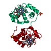 1yccS S: Starting model for refinement |
|---|---|
| Similar structure data |


-Links
 PDBj
PDBj


- Assembly
| Deposited unit | 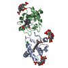
| ||||||||
|---|---|---|---|---|---|---|---|---|---|
| 1 | 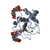
| ||||||||
| 2 | 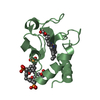
| ||||||||
| Unit cell |
|
-Components
| #1: Protein | Mass: 12041.770 Da / Num. of mol.: 2 / Mutation: T-5A, C102T Source method: isolated from a genetically manipulated source Source: (gene. exp.) Saccharomyces cerevisiae (strain ATCC 204508 / S288c) (yeast)Strain: ATCC 204508 / S288c / Gene: CYC1, YJR048W, J1653 / Production host:  Escherichia coli BL21(DE3) (bacteria) / References: UniProt: P00044 Escherichia coli BL21(DE3) (bacteria) / References: UniProt: P00044#2: Chemical | Heme C  Mass: 618.503 Da / Num. of mol.: 2 / Source method: obtained synthetically / Formula: C34H34FeN4O4 Mass: 618.503 Da / Num. of mol.: 2 / Source method: obtained synthetically / Formula: C34H34FeN4O4#3: Chemical | 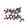  Mass: 800.514 Da / Num. of mol.: 3 / Source method: obtained synthetically / Formula: C32H36O16P4 Mass: 800.514 Da / Num. of mol.: 3 / Source method: obtained synthetically / Formula: C32H36O16P4#4: Chemical | ChemComp-CL / | Chloride  Mass: 35.453 Da / Num. of mol.: 1 / Source method: obtained synthetically / Formula: Cl Mass: 35.453 Da / Num. of mol.: 1 / Source method: obtained synthetically / Formula: Cl#5: Water | ChemComp-HOH / | Water Mass: 18.015 Da / Num. of mol.: 282 / Source method: isolated from a natural source / Formula: H2O Mass: 18.015 Da / Num. of mol.: 282 / Source method: isolated from a natural source / Formula: H2O |
|---|
-Experimental details
-Experiment
| Experiment | Method: X-RAY DIFFRACTION / Number of used crystals: 1 |
|---|
- Sample preparation
| Crystal | Density Matthews: 2.39 Å3/Da / Density % sol: 48.62 % |
|---|---|
| Crystal grow | Temperature: 293 K / Method: vapor diffusion, hanging drop / pH: 5.6 Details: 20 % PEG 8000, 50 mM NaCl, 50 mM sodium acetate (pH 5.6). [Cytochrome c] = 0.75 mM and [Methylphosphonatocalix[4]arene] = 0.3 mM |
-Data collection
| Diffraction | Mean temperature: 100 K |
|---|---|
| Diffraction source | Source: SYNCHROTRON / Site: APS  / Beamline: 24-ID-C / Wavelength: 0.979 Å / Beamline: 24-ID-C / Wavelength: 0.979 Å |
| Detector | Type: DECTRIS PILATUS 6M-F / Detector: PIXEL / Date: Aug 13, 2016 |
| Radiation | Protocol: SINGLE WAVELENGTH / Monochromatic (M) / Laue (L): M / Scattering type: x-ray |
| Radiation wavelength | Wavelength: 0.979 Å / Relative weight: 1 |
| Reflection | Resolution: 1.5→47.74 Å / Num. obs: 34591 / % possible obs: 99.2 % / Redundancy: 5.5 % / CC1/2: 0.999 / Rmerge(I) obs: 0.055 / Rpim(I) all: 0.026 / Net I/σ(I): 18.7 |
| Reflection shell | Resolution: 1.5→1.55 Å / Redundancy: 5.4 % / Rmerge(I) obs: 0.055 / Mean I/σ(I) obs: 5.1 / Num. unique obs: 3452 / Rpim(I) all: 0.158 / % possible all: 99.4 |
- Processing
| Software |
| ||||||||||||||||||||||||||||||||||||||||||||||||||||||||||||
|---|---|---|---|---|---|---|---|---|---|---|---|---|---|---|---|---|---|---|---|---|---|---|---|---|---|---|---|---|---|---|---|---|---|---|---|---|---|---|---|---|---|---|---|---|---|---|---|---|---|---|---|---|---|---|---|---|---|---|---|---|---|
| Refinement | Method to determine structure: MOLECULAR REPLACEMENT Starting model: 1YCC Resolution: 1.5→47.74 Å / Cor.coef. Fo:Fc: 0.97 / Cor.coef. Fo:Fc free: 0.961 / SU B: 1.649 / SU ML: 0.06 / Cross valid method: THROUGHOUT / σ(F): 0 / ESU R: 0.081 / ESU R Free: 0.081 Details: HYDROGENS HAVE BEEN ADDED IN THE RIDING POSITIONS U VALUES : REFINED INDIVIDUALLY
| ||||||||||||||||||||||||||||||||||||||||||||||||||||||||||||
| Solvent computation | Ion probe radii: 0.8 Å / Shrinkage radii: 0.8 Å / VDW probe radii: 1.2 Å | ||||||||||||||||||||||||||||||||||||||||||||||||||||||||||||
| Displacement parameters | Biso max: 143.7 Å2 / Biso mean: 20.624 Å2 / Biso min: 9.15 Å2
| ||||||||||||||||||||||||||||||||||||||||||||||||||||||||||||
| Refinement step | Cycle: final / Resolution: 1.5→47.74 Å
| ||||||||||||||||||||||||||||||||||||||||||||||||||||||||||||
| Refine LS restraints |
| ||||||||||||||||||||||||||||||||||||||||||||||||||||||||||||
| LS refinement shell | Resolution: 1.5→1.539 Å / Rfactor Rfree error: 0 / Total num. of bins used: 20
|
