 Movie
Movie Controller

[English] 日本語
 Yorodumi
- PDB-5n9a: Crystal Structure of Drosophila DHX36 helicase in complex with GT... -
+ Open data

- Basic information
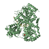
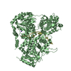
| Entry | Database: PDB / ID: 5n9a | ||||||
|---|---|---|---|---|---|---|---|
| Title | Crystal Structure of Drosophila DHX36 helicase in complex with GTTAGGGTT | ||||||
 Components Components |
| ||||||
 Keywords Keywords |  HYDROLASE / Helicase DExH ssDNA HYDROLASE / Helicase DExH ssDNA | ||||||
| Function / homology |  Function and homology information Function and homology informationDEx/H-box helicases activate type I IFN and inflammatory cytokines production / G-quadruplex DNA unwinding / G-quadruplex DNA binding / DNA helicase activity / helicase activity / G-quadruplex RNA binding / RNA helicase activity / RNA helicase / hydrolase activity / ATP binding ...DEx/H-box helicases activate type I IFN and inflammatory cytokines production / G-quadruplex DNA unwinding / G-quadruplex DNA binding / DNA helicase activity / helicase activity / G-quadruplex RNA binding / RNA helicase activity / RNA helicase / hydrolase activity / ATP binding / nucleus / cytoplasmSimilarity search - Function | ||||||
| Biological species |  Drosophila melanogaster (fruit fly) Drosophila melanogaster (fruit fly) Homo sapiens (human) Homo sapiens (human) | ||||||
| Method | X-RAY DIFFRACTION / SYNCHROTRON / MOLECULAR REPLACEMENT / Resolution: 3.036 Å | ||||||
 Authors Authors | Chen, W.-F. / Rety, S. / Guo, H.-L. / Wu, W.-Q. / Liu, N.-N. / Liu, Q.-W. / Dai, Y.-X. / Xi, X.-G. | ||||||
 Citation Citation | Journal: Structure / Year: 2018 Title: Molecular Mechanistic Insights into Drosophila DHX36-Mediated G-Quadruplex Unfolding: A Structure-Based Model. Authors: Chen, W.F. / Rety, S. / Guo, H.L. / Dai, Y.X. / Wu, W.Q. / Liu, N.N. / Auguin, D. / Liu, Q.W. / Hou, X.M. / Dou, S.X. / Xi, X.G. | ||||||
| History |
|
- Structure visualization
| Structure viewer | Molecule: MolmilJmol/JSmol |
|---|
- Downloads & links
-Download
| PDBx/mmCIF format | 5n9a.cif.gz | 715 KB | Display | PDBx/mmCIF format |
|---|---|---|---|---|
| PDB format | pdb5n9a.ent.gz | 590.4 KB | Display | PDB format |
| PDBx/mmJSON format | 5n9a.json.gz | Tree view | PDBx/mmJSON format | |
| Others |  Other downloads Other downloads |
-Validation report
| Arichive directory | https://data.pdbj.org/pub/pdb/validation_reports/n9/5n9aftp://data.pdbj.org/pub/pdb/validation_reports/n9/5n9a | HTTPS FTP |
|---|
-Related structure data
| Related structure data |  5n8rSC  5n8sC  5n90C  5n94C  5n96C  5n98C  5n9dC S: Starting model for refinement C: citing same article ( |
|---|---|
| Similar structure data |


-Links
 PDBj
PDBj- Assembly
| Deposited unit | 
| ||||||||
|---|---|---|---|---|---|---|---|---|---|
| 1 | 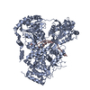
| ||||||||
| 2 | 
| ||||||||
| Unit cell |
|
-Components
| #1: Protein | Mass: 108401.367 Da / Num. of mol.: 2 Source method: isolated from a genetically manipulated source Source: (gene. exp.) Drosophila melanogaster (fruit fly) / Gene: CG9323, Dmel_CG9323 / Plasmid: pTwin1 / Production host:  Escherichia coli (E. coli) / Variant (production host): C2566H / References: UniProt: Q8SWT2, EC: 3.6.1.3 Escherichia coli (E. coli) / Variant (production host): C2566H / References: UniProt: Q8SWT2, EC: 3.6.1.3#2: DNA chain | Mass: 2801.844 Da / Num. of mol.: 2 / Source method: obtained synthetically / Source: (synth.) Homo sapiens (human)#3: Water | ChemComp-HOH / | Water Mass: 18.015 Da / Num. of mol.: 3 / Source method: isolated from a natural source / Formula: H2O Mass: 18.015 Da / Num. of mol.: 3 / Source method: isolated from a natural source / Formula: H2O |
|---|
-Experimental details
-Experiment
| Experiment | Method: X-RAY DIFFRACTION / Number of used crystals: 1 |
|---|
- Sample preparation
| Crystal | Density Matthews: 2.69 Å3/Da / Density % sol: 54.3 % |
|---|---|
| Crystal grow | Temperature: 298 K / Method: vapor diffusion, sitting drop / pH: 6.5 Details: 0.1M MES monohydrate-imidazole 20mM sodium formate 20mM ammonium acetate 20mM sodium citrate tribasic hydrate 20mM sodium oxamate 20mM potassium sodium tartrate tetrahydrate |
-Data collection
| Diffraction | Mean temperature: 100 K |
|---|---|
| Diffraction source | Source: SYNCHROTRON / Site: SSRF  / Beamline: BL17U / Wavelength: 0.9791 Å / Beamline: BL17U / Wavelength: 0.9791 Å |
| Detector | Type: ADSC QUANTUM 315r / Detector: CCD / Date: Jul 17, 2016 |
| Radiation | Protocol: SINGLE WAVELENGTH / Monochromatic (M) / Laue (L): M / Scattering type: x-ray |
| Radiation wavelength | Wavelength: 0.9791 Å / Relative weight: 1 |
| Reflection | Resolution: 3.036→69.27 Å / Num. obs: 44188 / % possible obs: 96.04 % / Observed criterion σ(I): 2 / Redundancy: 3.7 % / Biso Wilson estimate: 62.33 Å2 / CC1/2: 0.991 / Rmerge(I) obs: 0.1171 / Net I/σ(I): 9.18 |
| Reflection shell | Resolution: 3.036→3.145 Å / Redundancy: 3.8 % / Rmerge(I) obs: 0.5895 / Mean I/σ(I) obs: 2.34 / Num. unique obs: 4476 / CC1/2: 0.731 / % possible all: 99.89 |
- Processing
| Software |
| ||||||||||||||||||||||||||||||||||||||||||||||||||||||||||||||||||||||||||||||||||||||||||||||||||||||||||||||||||||||||||||||||||||||||||||||||||||||||||||||||||||||||||||||||||||||||||||||||||||||||||||||||||||||||||||||||||||||||||||||||||||||||||
|---|---|---|---|---|---|---|---|---|---|---|---|---|---|---|---|---|---|---|---|---|---|---|---|---|---|---|---|---|---|---|---|---|---|---|---|---|---|---|---|---|---|---|---|---|---|---|---|---|---|---|---|---|---|---|---|---|---|---|---|---|---|---|---|---|---|---|---|---|---|---|---|---|---|---|---|---|---|---|---|---|---|---|---|---|---|---|---|---|---|---|---|---|---|---|---|---|---|---|---|---|---|---|---|---|---|---|---|---|---|---|---|---|---|---|---|---|---|---|---|---|---|---|---|---|---|---|---|---|---|---|---|---|---|---|---|---|---|---|---|---|---|---|---|---|---|---|---|---|---|---|---|---|---|---|---|---|---|---|---|---|---|---|---|---|---|---|---|---|---|---|---|---|---|---|---|---|---|---|---|---|---|---|---|---|---|---|---|---|---|---|---|---|---|---|---|---|---|---|---|---|---|---|---|---|---|---|---|---|---|---|---|---|---|---|---|---|---|---|---|---|---|---|---|---|---|---|---|---|---|---|---|---|---|---|---|---|---|---|---|---|---|---|---|---|---|---|---|---|---|---|---|
| Refinement | Method to determine structure: MOLECULAR REPLACEMENT Starting model: 5N8R Resolution: 3.036→69.27 Å / SU ML: 0.56 / Cross valid method: FREE R-VALUE / σ(F): 1.35 / Phase error: 34.22
| ||||||||||||||||||||||||||||||||||||||||||||||||||||||||||||||||||||||||||||||||||||||||||||||||||||||||||||||||||||||||||||||||||||||||||||||||||||||||||||||||||||||||||||||||||||||||||||||||||||||||||||||||||||||||||||||||||||||||||||||||||||||||||
| Solvent computation | Shrinkage radii: 0.9 Å / VDW probe radii: 1.11 Å | ||||||||||||||||||||||||||||||||||||||||||||||||||||||||||||||||||||||||||||||||||||||||||||||||||||||||||||||||||||||||||||||||||||||||||||||||||||||||||||||||||||||||||||||||||||||||||||||||||||||||||||||||||||||||||||||||||||||||||||||||||||||||||
| Refinement step | Cycle: LAST / Resolution: 3.036→69.27 Å
| ||||||||||||||||||||||||||||||||||||||||||||||||||||||||||||||||||||||||||||||||||||||||||||||||||||||||||||||||||||||||||||||||||||||||||||||||||||||||||||||||||||||||||||||||||||||||||||||||||||||||||||||||||||||||||||||||||||||||||||||||||||||||||
| Refine LS restraints |
| ||||||||||||||||||||||||||||||||||||||||||||||||||||||||||||||||||||||||||||||||||||||||||||||||||||||||||||||||||||||||||||||||||||||||||||||||||||||||||||||||||||||||||||||||||||||||||||||||||||||||||||||||||||||||||||||||||||||||||||||||||||||||||
| LS refinement shell |
| ||||||||||||||||||||||||||||||||||||||||||||||||||||||||||||||||||||||||||||||||||||||||||||||||||||||||||||||||||||||||||||||||||||||||||||||||||||||||||||||||||||||||||||||||||||||||||||||||||||||||||||||||||||||||||||||||||||||||||||||||||||||||||
| Refinement TLS params. | Method: refined / Refine-ID: X-RAY DIFFRACTION
| ||||||||||||||||||||||||||||||||||||||||||||||||||||||||||||||||||||||||||||||||||||||||||||||||||||||||||||||||||||||||||||||||||||||||||||||||||||||||||||||||||||||||||||||||||||||||||||||||||||||||||||||||||||||||||||||||||||||||||||||||||||||||||
| Refinement TLS group |
|
