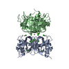
[English] 日本語
Yorodumi
- PDB-5mwv: Solid-state NMR Structure of outer membrane protein G in lipid bi... -

+
Open data


ID or keywords:

Loading...

-
Basic information

Entry
Database: PDB / ID: 5mwv
TitleSolid-state NMR Structure of outer membrane protein G in lipid bilayers
ComponentsOuter membrane protein G
KeywordsMEMBRANE PROTEIN / porin beta-barrel membrane lipid
Function / homology
Function and homology information


carbohydrate transmembrane transport / oligosaccharide transporting porin activity / maltose transporting porin activity / porin activity / pore complex / monoatomic ion transport / cell outer membrane
Similarity search - Function
Outer membrane porin G / Outer membrane protein G (OmpG)
Similarity search - Domain/homology
Outer membrane porin G
Similarity search - Component
Biological speciesEscherichia coli K12 (bacteria)
MethodSOLID-STATE NMR / molecular dynamics
AuthorsRetel, J.S. / Nieuwkoop, A.J. / Hiller, M. / Higman, V.A. / Barbet-Massin, E. / Stanek, J. / Andreas, L.B. / Franks, W.T. / van Rossum, B.-J. / Vinothkumar, K.R. ...Retel, J.S. / Nieuwkoop, A.J. / Hiller, M. / Higman, V.A. / Barbet-Massin, E. / Stanek, J. / Andreas, L.B. / Franks, W.T. / van Rossum, B.-J. / Vinothkumar, K.R. / Handel, L. / de Palma, G.G. / Bardiaux, B. / Pintacuda, G. / Emsley, L. / Kuelbrandt, W. / Oschkinat, H.
Funding support Germany, 4items
OrganizationGrant numberCountry
European UnionBioNMR / 261863 Germany
European UnioniNext / GA 653706
German Research FoundationSFB 740 Germany
German Research FoundationOS106/9 Germany
CitationJournal: Nat Commun / Year: 2017
Title: Structure of outer membrane protein G in lipid bilayers.
Authors: Retel, J.S. / Nieuwkoop, A.J. / Hiller, M. / Higman, V.A. / Barbet-Massin, E. / Stanek, J. / Andreas, L.B. / Franks, W.T. / van Rossum, B.J. / Vinothkumar, K.R. / Handel, L. / de Palma, G.G. ...Authors: Retel, J.S. / Nieuwkoop, A.J. / Hiller, M. / Higman, V.A. / Barbet-Massin, E. / Stanek, J. / Andreas, L.B. / Franks, W.T. / van Rossum, B.J. / Vinothkumar, K.R. / Handel, L. / de Palma, G.G. / Bardiaux, B. / Pintacuda, G. / Emsley, L. / Kuhlbrandt, W. / Oschkinat, H.
History
DepositionJan 20, 2017Deposition site: PDBE / Processing site: PDBE
Revision 1.0Dec 27, 2017Provider: repository / Type: Initial release
Revision 1.1Aug 21, 2019Group: Data collection / Category: pdbx_nmr_software / Item: _pdbx_nmr_software.name
Revision 1.2May 15, 2024Group: Data collection / Database references
Category: chem_comp_atom / chem_comp_bond ...chem_comp_atom / chem_comp_bond / database_2 / pdbx_nmr_spectrometer
Item: _database_2.pdbx_DOI / _database_2.pdbx_database_accession / _pdbx_nmr_spectrometer.model

-
Structure visualization

Structure viewerMolecule:
MolmilJmol/JSmol

Downloads & links

-
Assembly

Deposited unit
A: Outer membrane protein G


Theoretical massNumber of molelcules
Total (without water)32,9371
Polymers32,9371
Non-polymers00
Water0
1


  • Idetical with deposited unit
  • defined by author&software
TypeNameSymmetry operationNumber
identity operation1_5551
Buried area0 Å2
ΔGint0 kcal/mol
Surface area22310 Å2
MethodPISA
NMR ensembles
DataCriteria
Number of conformers (submitted / calculated)15 / 200structures with the lowest energy
RepresentativeModel #1lowest energy

-
Components

#1: Protein Outer membrane protein G /


Mass: 32936.527 Da / Num. of mol.: 1
Source method: isolated from a genetically manipulated source
Source: (gene. exp.) Escherichia coli K12 (bacteria) / Gene: ompG, b1319, JW1312 / Production host: Escherichia coli (E. coli) / References: UniProt: P76045

-
Experimental details

-
Experiment

ExperimentMethod: SOLID-STATE NMR
NMR experiment
Conditions-IDExperiment-IDSolution-IDSample stateSpectrometer-IDType
111isotropic23D (H)N(HH)NH RFDR 2ms
121isotropic23D (H)NHH RFDR 2ms
232isotropic12D 13C-13C DARR 200ms
242isotropic12D 13C-13C DARR 400ms
253isotropic12D 13C-13C DARR 200ms
263isotropic12D 13C-13C DARR 400ms
284isotropic12D 13C-13C DARR 150ms
274isotropic12D 13C-13C DARR 400ms
295isotropic12D 13C-13C DARR 150ms
2105isotropic12D 13C-13C DARR 400ms
2116isotropic12D 13C-13C DARR 150ms
2126isotropic12D 13C-13C DARR 400ms
2137isotropic12D 13C-13C DARR 500ms
1141isotropic23D (H)CANH
3158isotropic33D (H)CANH
3168isotropic33D (HCO)CA(CO)NH
3178isotropic33D (HCA)CB(CA)NH
3188isotropic33D (HCA)CBCA(CO)NH
3198isotropic33D (H)CONH
3208isotropic33D (H)CO(CA)NH

-
Sample preparation

Details
TypeSolution-IDContentsDetailsLabelSolvent system
solid10.66 w/w [U-13C; U-15N; U-2H] backexchanged 1H Outer Membrane Protein G, 0.33 w/w lipids, MES BufferFully deuterated. Subsequently the exchangeable sites were back-exchanged with 100% 1H.deuterated-back-exchanged-OmpGMES Buffer
solid80.66 w/w [U-13C; U-15N; U-2H] backexchanged 1H Outer Membrane Protein G, HEPES BufferFully deuterated. Subsequently the exchangeable sites were back-exchanged with 70% 1H.deuterated-70-back-exchanged-OmpGHEPES Buffer
solid20.66 w/w [U-15N; U-1H] Outer Membrane Protein G, 0.33 w/w lipids, HEPES BufferUniformly 1H and 15N labeled. 13C labeling as obtained by using 1,3-labeled glycerol as the sole carbon source during protein expression1,3-glycerol-OmpGHEPES Buffer
solid30.66 w/w [U-15N; U-1H] Outer Membrane Protein G, 0.33 w/w lipids, HEPES BufferUniformly 1H and 15N labeled. 13C labeling as obtained by using 2-labeled glycerol as the sole carbon source during protein expression2-glycerol-OmpGHEPES Buffer
solid40.66 w/w [U-15N; U-1H] Outer Membrane Protein G, 0.33 w/w lipids, HEPES BufferUniformly 1H and 15N labeled. The residues T,E,M,P,Q,A,N,S,G are 13C labeled as obtained by using 1,3-labeled glycerol as the sole carbon source during protein expression. Other amino acids are not 13C labeled.1,3-TEMPQANDSG-OmpGHEPES Buffer
solid50.66 w/w [U-15N; U-1H] Outer Membrane Protein G, 0.33 w/w lipids, HEPES BufferUniformly 1H and 15N labeled. The residues T,E,M,P,Q,A,N,S,G are 13C labeled as obtained by using 2-labeled glycerol as the sole carbon source during protein expression. Other amino acids are not 13C labeled.2-TEMPQANDSG-OmpGHEPES Buffer
solid60.66 w/w [U-15N; U-1H] Outer Membrane Protein G, 0.33 w/w na lipids, HEPES BufferUniformly 1H and 15N labeled. The residues S,H,L,Y,G,W,A,F,V are 13C labeled as obtained by using 2-labeled glycerol as the sole carbon source during protein expression. Other amino acids are not 13C labeled.2-SHLYGWAFV-OmpGHEPES Buffer
solid70.66 w/w [U-15N; U-1H] Outer Membrane Protein G, 0.33 w/w lipids, HEPES BufferUniformly 1H and 15N labeled. The residues G and A are 13C labeled. F and Y are 13C labeled on the CA and CB.GAF23Y23-OmpGHEPES Buffer
Sample
Conc. (mg/ml)ComponentIsotopic labelingSolution-ID
0.66 w/wOuter Membrane Protein G[U-13C; U-15N; U-2H] backexchanged 1H1
0.33 w/wlipidsnatural abundance1
0.66 w/wOuter Membrane Protein G[U-13C; U-15N; U-2H] backexchanged 1H8
0.66 w/wOuter Membrane Protein G[U-15N; U-1H]2
0.33 w/wlipidsnatural abundance2
0.66 w/wOuter Membrane Protein G[U-15N; U-1H]3
0.33 w/wlipidsnatural abundance3
0.66 w/wOuter Membrane Protein G[U-15N; U-1H]4
0.33 w/wlipidsnatural abundance4
0.66 w/wOuter Membrane Protein G[U-15N; U-1H]5
0.33 w/wlipidsnatural abundance5
0.66 w/wOuter Membrane Protein G[U-15N; U-1H]6
0.33 w/wlipidsna6
0.66 w/wOuter Membrane Protein G[U-15N; U-1H]7
0.33 w/wlipidsnatural abundance7
Sample conditions

Ionic strength: 50 mM NaCl mM / Pressure: 1 atm / Temperature: 280 K

Conditions-IDDetailsLabelpHTemperature err
1There is a large error on the sample temperature due to fast MAS spinning (60 kHz).proton-detection-distances6.3 20
3There is a large error on the sample temperature due to fast MAS spinning (60kHz).proton-detection-initial-assignment6.8 20
2Condition at experiments at lower MAS rates (13 kHz).carbon-detection6.8

-
NMR measurement

NMR spectrometer
TypeManufacturerModelField strength (MHz)Spectrometer-IDDetails
Bruker AVANCEBrukerAVANCE9001Berlin
Bruker AVANCEBrukerAVANCE10002Lyon
Bruker AVANCEBrukerAVANCE8003Lyon

-
Processing

NMR software
NameVersionDeveloperClassification
CNSBrunger A. T. et.al.refinement
ARIA1.21Linge, O'Donoghue and Nilgesstructure calculation
CcpNmr Analysis2.4CCPNchemical shift assignment
CcpNmr Analysis2.4CCPNpeak picking
TALOSTALOS+Cornilescu, Delaglio and Baxdata analysis
RefinementMethod: molecular dynamics / Software ordinal: 1
NMR representativeSelection criteria: lowest energy
NMR ensembleConformer selection criteria: structures with the lowest energy
Conformers calculated total number: 200 / Conformers submitted total number: 15

+
About Yorodumi

-
News

-
Feb 9, 2022. New format data for meta-information of EMDB entries

New format data for meta-information of EMDB entries

  • Version 3 of the EMDB header file is now the official format.
  • The previous official version 1.9 will be removed from the archive.

Related info.:EMDB header

External links:wwPDB to switch to version 3 of the EMDB data model

-
Aug 12, 2020. Covid-19 info

Covid-19 info

URL: https://pdbj.org/emnavi/covid19.php

New page: Covid-19 featured information page in EM Navigator.

Related info.:Covid-19 info / Mar 5, 2020. Novel coronavirus structure data

+
Mar 5, 2020. Novel coronavirus structure data

Novel coronavirus structure data

Related info.:Yorodumi Speices / Aug 12, 2020. Covid-19 info

External links:COVID-19 featured content - PDBj / Molecule of the Month (242):Coronavirus Proteases

+
Jan 31, 2019. EMDB accession codes are about to change! (news from PDBe EMDB page)

EMDB accession codes are about to change! (news from PDBe EMDB page)

  • The allocation of 4 digits for EMDB accession codes will soon come to an end. Whilst these codes will remain in use, new EMDB accession codes will include an additional digit and will expand incrementally as the available range of codes is exhausted. The current 4-digit format prefixed with “EMD-” (i.e. EMD-XXXX) will advance to a 5-digit format (i.e. EMD-XXXXX), and so on. It is currently estimated that the 4-digit codes will be depleted around Spring 2019, at which point the 5-digit format will come into force.
  • The EM Navigator/Yorodumi systems omit the EMD- prefix.

Related info.:Q: What is EMD? / ID/Accession-code notation in Yorodumi/EM Navigator

External links:EMDB Accession Codes are Changing Soon! / Contact to PDBj

+
Jul 12, 2017. Major update of PDB

Major update of PDB

  • wwPDB released updated PDB data conforming to the new PDBx/mmCIF dictionary.
  • This is a major update changing the version number from 4 to 5, and with Remediation, in which all the entries are updated.
  • In this update, many items about electron microscopy experimental information are reorganized (e.g. em_software).
  • Now, EM Navigator and Yorodumi are based on the updated data.

External links:wwPDB Remediation / Enriched Model Files Conforming to OneDep Data Standards Now Available in the PDB FTP Archive

-
Yorodumi

Thousand views of thousand structures

  • Yorodumi is a browser for structure data from EMDB, PDB, SASBDB, etc.
  • This page is also the successor to EM Navigator detail page, and also detail information page/front-end page for Omokage search.
  • The word "yorodu" (or yorozu) is an old Japanese word meaning "ten thousand". "mi" (miru) is to see.

Related info.:EMDB / PDB / SASBDB / Comparison of 3 databanks / Yorodumi Search / Aug 31, 2016. New EM Navigator & Yorodumi / Yorodumi Papers / Jmol/JSmol / Function and homology information / Changes in new EM Navigator and Yorodumi

Read more