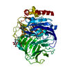
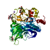
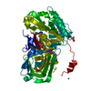
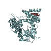
- PDB-5mwm: INOSITOL 1,3,4,5,6-PENTAKISPHOSPHATE 2-KINASE FROM M. MUSCULUS IN... -
+
Open data
ID or keywords:
Loading...
-
Basic information
Entry
Database: PDB / ID: 5mwm
Title
INOSITOL 1,3,4,5,6-PENTAKISPHOSPHATE 2-KINASE FROM M. MUSCULUS IN COMPLEX WITH IP6
Components
Inositol-pentakisphosphate 2-kinase
Keywords
TRANSFERASE / protein structure / mammal IPK / inositol kinases
Function / homology
Function and homology information
Synthesis of IPs in the nucleus / Synthesis of pyrophosphates in the cytosol / inositol-pentakisphosphate 2-kinase / inositol pentakisphosphate 2-kinase activity / inositol phosphate biosynthetic process / phosphorylation / ATP binding / nucleus / cytoplasm Similarity search - Function
Resolution: 2.6→57.46 Å / Cor.coef. Fo:Fc: 0.94 / Cor.coef. Fo:Fc free: 0.919 / SU B: 39.012 / SU ML: 0.67 / Cross valid method: THROUGHOUT / ESU R: 1.229 / ESU R Free: 0.37 / Details: HYDROGENS HAVE BEEN ADDED IN THE RIDING POSITIONS
Rfactor
Num. reflection
% reflection
Selection details
Rfree
0.28954
707
4.7 %
RANDOM
Rwork
0.25343
-
-
-
obs
0.25523
14393
98.99 %
-
Solvent computation
Ion probe radii: 0.8 Å / Shrinkage radii: 0.8 Å / VDW probe radii: 1.2 Å
 Movie
Movie Controller

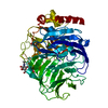
 Yorodumi
 Open data
 Basic information
 Components
 Keywords
 Function and homology information
 Authors
 Spain, 1items
  Citation
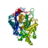
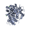
 Structure visualization
 Downloads & links
 Other downloads


 PDBj
PDBj


 Assembly
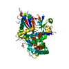


 Mass: 660.035 Da / Num. of mol.: 1 / Source method: obtained synthetically / Formula: C6H18O24P6
 Mass: 65.409 Da / Num. of mol.: 1 / Source method: obtained synthetically / Formula: Zn
 Mass: 18.015 Da / Num. of mol.: 26 / Source method: isolated from a natural source / Formula: H2O
 Sample preparation
 Processing