 Movie
Movie Controller


+ Open data

- Basic information
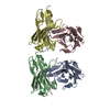
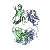
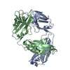
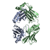
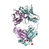
| Entry | Database: PDB / ID: 5mtl | ||||||
|---|---|---|---|---|---|---|---|
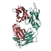
| Title | Crystal structure of an amyloidogenic light chain | ||||||
 Components Components | light chain dimer,IGL@ protein,IGL@ protein | ||||||
 Keywords Keywords |  IMMUNE SYSTEM / light chain dimer / light chain amyloidosis / immunoglobulin fold / protein aggregation IMMUNE SYSTEM / light chain dimer / light chain amyloidosis / immunoglobulin fold / protein aggregation | ||||||
| Function / homology |  Function and homology information Function and homology information | ||||||
| Biological species |  Homo sapiens (human) Homo sapiens (human) | ||||||
| Method | X-RAY DIFFRACTION / SYNCHROTRON / MOLECULAR REPLACEMENT / Resolution: 2.45 Å | ||||||
 Authors Authors | Oberti, L. / Rognoni, P. / Russo, R. / Bacarizo, J. / Bolognesi, M. / Ricagno, S. | ||||||
 Citation Citation | Journal: Sci Rep / Year: 2017 Title: Concurrent structural and biophysical traits link with immunoglobulin light chains amyloid propensity. Authors: Oberti, L. / Rognoni, P. / Barbiroli, A. / Lavatelli, F. / Russo, R. / Maritan, M. / Palladini, G. / Bolognesi, M. / Merlini, G. / Ricagno, S. | ||||||
| History |
|
- Structure visualization
| Structure viewer | Molecule: MolmilJmol/JSmol |
|---|
- Downloads & links
-Download
| PDBx/mmCIF format | 5mtl.cif.gz | 156.6 KB | Display | PDBx/mmCIF format |
|---|---|---|---|---|
| PDB format | pdb5mtl.ent.gz | 123.8 KB | Display | PDB format |
| PDBx/mmJSON format | 5mtl.json.gz | Tree view | PDBx/mmJSON format | |
| Others |  Other downloads Other downloads |
-Validation report
| Arichive directory | https://data.pdbj.org/pub/pdb/validation_reports/mt/5mtlftp://data.pdbj.org/pub/pdb/validation_reports/mt/5mtl | HTTPS FTP |
|---|
-Related structure data
| Related structure data | 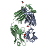 5m6aC  5m6iSC  5m76C 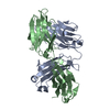 5mudC 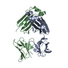 5muhC  5mvgC S: Starting model for refinement C: citing same article ( |
|---|---|
| Similar structure data |


-Links
 PDBj
PDBj

- Assembly
| Deposited unit | 
| ||||||||
|---|---|---|---|---|---|---|---|---|---|
| 1 | 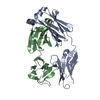
| ||||||||
| 2 | 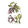
| ||||||||
| Unit cell |
|
-Components
| #1: Antibody | Mass: 22756.051 Da / Num. of mol.: 4 Source method: isolated from a genetically manipulated source Source: (gene. exp.) Homo sapiens (human) / Gene: IGL@ / Production host:  Escherichia coli (E. coli) / References: UniProt: Q567P1 Escherichia coli (E. coli) / References: UniProt: Q567P1#2: Water | ChemComp-HOH / | Water Mass: 18.015 Da / Num. of mol.: 44 / Source method: isolated from a natural source / Formula: H2O Mass: 18.015 Da / Num. of mol.: 44 / Source method: isolated from a natural source / Formula: H2O |
|---|
-Experimental details
-Experiment
| Experiment | Method: X-RAY DIFFRACTION / Number of used crystals: 1 |
|---|
- Sample preparation
| Crystal | Density Matthews: 2.88 Å3/Da / Density % sol: 57.27 % |
|---|---|
| Crystal grow | Temperature: 293 K / Method: vapor diffusion, sitting drop / pH: 6.5 / Details: Sodium cacodylate, 27% w/v PEG 2000 MME |
-Data collection
| Diffraction | Mean temperature: 126.15 K |
|---|---|
| Diffraction source | Source: SYNCHROTRON / Site: ESRF  / Beamline: ID23-2 / Wavelength: 0.873 Å / Beamline: ID23-2 / Wavelength: 0.873 Å |
| Detector | Type: DECTRIS PILATUS 2M-F / Detector: PIXEL / Date: Jun 15, 2014 |
| Radiation | Protocol: SINGLE WAVELENGTH / Monochromatic (M) / Laue (L): M / Scattering type: x-ray |
| Radiation wavelength | Wavelength: 0.873 Å / Relative weight: 1 |
| Reflection | Resolution: 2.45→28.57 Å / Num. obs: 36581 / % possible obs: 98.2 % / Redundancy: 3.3 % / Rmerge(I) obs: 0.126 / Net I/σ(I): 8.8 |
| Reflection shell | Resolution: 2.45→2.54 Å / Redundancy: 3.2 % / Rmerge(I) obs: 0.61 / Mean I/σ(I) obs: 2.2 / Num. unique obs: 3606 / % possible all: 97.9 |
- Processing
| Software |
| ||||||||||||||||||||||||||||||||||||||||||||||||||||||||||||||||||||||||||||||||||||||||||||||||||
|---|---|---|---|---|---|---|---|---|---|---|---|---|---|---|---|---|---|---|---|---|---|---|---|---|---|---|---|---|---|---|---|---|---|---|---|---|---|---|---|---|---|---|---|---|---|---|---|---|---|---|---|---|---|---|---|---|---|---|---|---|---|---|---|---|---|---|---|---|---|---|---|---|---|---|---|---|---|---|---|---|---|---|---|---|---|---|---|---|---|---|---|---|---|---|---|---|---|---|---|
| Refinement | Method to determine structure: MOLECULAR REPLACEMENT Starting model: 5M6I Resolution: 2.45→29.289 Å / SU ML: 0.37 / Cross valid method: FREE R-VALUE / σ(F): 1.98 / Phase error: 31.73 / Stereochemistry target values: ML
| ||||||||||||||||||||||||||||||||||||||||||||||||||||||||||||||||||||||||||||||||||||||||||||||||||
| Solvent computation | Shrinkage radii: 0.9 Å / VDW probe radii: 1.11 Å / Solvent model: FLAT BULK SOLVENT MODEL | ||||||||||||||||||||||||||||||||||||||||||||||||||||||||||||||||||||||||||||||||||||||||||||||||||
| Refinement step | Cycle: LAST / Resolution: 2.45→29.289 Å
| ||||||||||||||||||||||||||||||||||||||||||||||||||||||||||||||||||||||||||||||||||||||||||||||||||
| Refine LS restraints |
| ||||||||||||||||||||||||||||||||||||||||||||||||||||||||||||||||||||||||||||||||||||||||||||||||||
| LS refinement shell |
|
