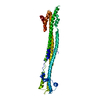
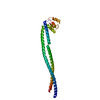
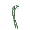
- PDB-5mq4: Crystal Structure of the leucine zipper of human PRKCBP1 -
+
Open data
ID or keywords:
Loading...
-
Basic information
Entry
Database: PDB / ID: 5mq4
Title
Crystal Structure of the leucine zipper of human PRKCBP1
Components
Protein kinase C-binding protein 1
Keywords
TRANSFERASE / Leucine zipper
Function / homology
Function and homology information
regulation of postsynaptic density protein 95 clustering / positive regulation of filopodium assembly / positive regulation of dendritic spine development / modulation of excitatory postsynaptic potential / site of DNA damage / positive regulation of dendritic spine maintenance / protein localization to chromatin / methylated histone binding / negative regulation of cell migration / dendritic shaft ...regulation of postsynaptic density protein 95 clustering / positive regulation of filopodium assembly / positive regulation of dendritic spine development / modulation of excitatory postsynaptic potential / site of DNA damage / positive regulation of dendritic spine maintenance / protein localization to chromatin / methylated histone binding / negative regulation of cell migration / dendritic shaft / positive regulation of transcription elongation by RNA polymerase II / double-strand break repair via homologous recombination / lysine-acetylated histone binding / transcription corepressor activity / nervous system development / chromatin organization / DNA-binding transcription factor binding / dendritic spine / protein domain specific binding / chromatin / nucleolus / negative regulation of transcription by RNA polymerase II / zinc ion binding / nucleoplasm / nucleus / cytoplasm Similarity search - Function
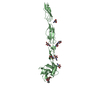
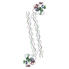
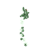
A: Protein kinase C-binding protein 1 B: Protein kinase C-binding protein 1 C: Protein kinase C-binding protein 1 D: Protein kinase C-binding protein 1 E: Protein kinase C-binding protein 1 F: Protein kinase C-binding protein 1 hetero molecules
In the structure databanks used in Yorodumi, some data are registered as the other names, "COVID-19 virus" and "2019-nCoV". Here are the details of the virus and the list of structure data.
Jan 31, 2019. EMDB accession codes are about to change! (news from PDBe EMDB page)
EMDB accession codes are about to change! (news from PDBe EMDB page)
The allocation of 4 digits for EMDB accession codes will soon come to an end. Whilst these codes will remain in use, new EMDB accession codes will include an additional digit and will expand incrementally as the available range of codes is exhausted. The current 4-digit format prefixed with “EMD-” (i.e. EMD-XXXX) will advance to a 5-digit format (i.e. EMD-XXXXX), and so on. It is currently estimated that the 4-digit codes will be depleted around Spring 2019, at which point the 5-digit format will come into force.
The EM Navigator/Yorodumi systems omit the EMD- prefix.
Related info.:Q: What is EMD? / ID/Accession-code notation in Yorodumi/EM Navigator
Yorodumi is a browser for structure data from EMDB, PDB, SASBDB, etc.
This page is also the successor to EM Navigator detail page, and also detail information page/front-end page for Omokage search.
The word "yorodu" (or yorozu) is an old Japanese word meaning "ten thousand". "mi" (miru) is to see.
Related info.:EMDB / PDB / SASBDB / Comparison of 3 databanks / Yorodumi Search / Aug 31, 2016. New EM Navigator & Yorodumi / Yorodumi Papers / Jmol/JSmol / Function and homology information / Changes in new EM Navigator and Yorodumi
 Movie
Movie Controller


 Open data
 Basic information
 Components
 Keywords
 TRANSFERASE /
  Function and homology information
 Authors
 Citation
 Structure visualization
 Downloads & links
 Other downloads
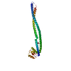


 PDBj
PDBj

 Assembly


 Mass: 96.063 Da / Num. of mol.: 5 / Source method: obtained synthetically / Formula: SO4
 Mass: 65.409 Da / Num. of mol.: 12 / Source method: obtained synthetically / Formula: Zn
 Mass: 18.015 Da / Num. of mol.: 1 / Source method: isolated from a natural source / Formula: H2O
 Sample preparation
 / Beamline: I02 / Wavelength: 0.97949 Å
 Processing