 Movie
Movie Controller

[English] 日本語
 Yorodumi
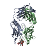
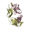
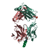
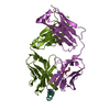
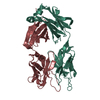
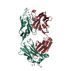
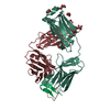
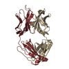
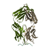
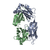
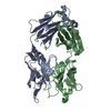
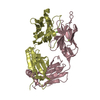
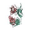
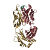
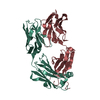
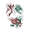
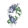
- PDB-5mo3: Crystal structure of DC8E8 Fab in the complex with a 14-mer tau p... -
+ Open data

- Basic information
| Entry | Database: PDB / ID: 5mo3 | ||||||||||||
|---|---|---|---|---|---|---|---|---|---|---|---|---|---|
| Title | Crystal structure of DC8E8 Fab in the complex with a 14-mer tau peptide at pH 8.5 | ||||||||||||
 Components Components |
| ||||||||||||
 Keywords Keywords |  IMMUNE SYSTEM / Fab / complex IMMUNE SYSTEM / Fab / complex | ||||||||||||
| Function / homology |  Function and homology information Function and homology informationplus-end-directed organelle transport along microtubule / axonal transport / histone-dependent DNA binding / neurofibrillary tangle assembly / positive regulation of diacylglycerol kinase activity / negative regulation of establishment of protein localization to mitochondrion / neurofibrillary tangle / positive regulation of protein localization to synapse / microtubule lateral binding / tubulin complex ...plus-end-directed organelle transport along microtubule / axonal transport / histone-dependent DNA binding / neurofibrillary tangle assembly / positive regulation of diacylglycerol kinase activity / negative regulation of establishment of protein localization to mitochondrion / neurofibrillary tangle / positive regulation of protein localization to synapse / microtubule lateral binding / tubulin complex / phosphatidylinositol bisphosphate binding / main axon / regulation of long-term synaptic depression / negative regulation of kinase activity / negative regulation of tubulin deacetylation / generation of neurons / regulation of chromosome organization / positive regulation of protein localization / rRNA metabolic process / internal protein amino acid acetylation / regulation of mitochondrial fission / intracellular distribution of mitochondria / axonal transport of mitochondrion / axon development / central nervous system neuron development / regulation of microtubule polymerization / microtubule polymerization / minor groove of adenine-thymine-rich DNA binding / lipoprotein particle binding / dynactin binding / glial cell projection / negative regulation of mitochondrial membrane potential / apolipoprotein binding / protein polymerization / negative regulation of mitochondrial fission / axolemma / Caspase-mediated cleavage of cytoskeletal proteins / regulation of microtubule polymerization or depolymerization / positive regulation of axon extension / Activation of AMPK downstream of NMDARs / regulation of microtubule cytoskeleton organization / supramolecular fiber organization / stress granule assembly / regulation of cellular response to heat / cytoplasmic microtubule organization / regulation of calcium-mediated signaling / positive regulation of microtubule polymerization / axon cytoplasm / somatodendritic compartment / cellular response to brain-derived neurotrophic factor stimulus / synapse assembly / phosphatidylinositol binding / nuclear periphery / cellular response to nerve growth factor stimulus / positive regulation of superoxide anion generation / protein phosphatase 2A binding / regulation of autophagy / astrocyte activation / response to lead ion / synapse organization / microglial cell activation / Hsp90 protein binding / regulation of synaptic plasticity / PKR-mediated signaling / protein homooligomerization / memory / cellular response to reactive oxygen species / microtubule cytoskeleton organization / SH3 domain binding / cytoplasmic ribonucleoprotein granule / activation of cysteine-type endopeptidase activity involved in apoptotic process / neuron projection development / microtubule cytoskeleton / cell-cell signaling / protein-macromolecule adaptor activity / actin binding / single-stranded DNA binding / cellular response to heat / cell body / protein-folding chaperone binding / growth cone / microtubule binding / double-stranded DNA binding / microtubule / sequence-specific DNA binding / amyloid fibril formation / dendritic spine / learning or memory / nuclear speck / neuron projection / membrane raft / axon / negative regulation of gene expression / neuronal cell body / DNA damage response / dendrite / protein kinase binding / enzyme binding / mitochondrion / DNA bindingSimilarity search - Function | ||||||||||||
| Biological species |  Mus musculus (house mouse) Mus musculus (house mouse) Homo sapiens (human) Homo sapiens (human) | ||||||||||||
| Method | X-RAY DIFFRACTION / SYNCHROTRON / MOLECULAR REPLACEMENT / molecular replacement / Resolution: 1.69 Å | ||||||||||||
 Authors Authors | Skrabana, R. / Novak, M. | ||||||||||||
 Citation Citation | Journal: To be published Title: Crystal structure of DC8E8 Fab in the complex with a 14-mer tau peptide at pH 8.5 Authors: Skrabana, R. / Novak, M. | ||||||||||||
| History |
|
- Structure visualization
| Structure viewer | Molecule: MolmilJmol/JSmol |
|---|
- Downloads & links
-Download
| PDBx/mmCIF format | 5mo3.cif.gz | 112.9 KB | Display | PDBx/mmCIF format |
|---|---|---|---|---|
| PDB format | pdb5mo3.ent.gz | 84.9 KB | Display | PDB format |
| PDBx/mmJSON format | 5mo3.json.gz | Tree view | PDBx/mmJSON format | |
| Others |  Other downloads Other downloads |
-Validation report
| Arichive directory | https://data.pdbj.org/pub/pdb/validation_reports/mo/5mo3ftp://data.pdbj.org/pub/pdb/validation_reports/mo/5mo3 | HTTPS FTP |
|---|
-Related structure data
| Related structure data |  4oz4 S: Starting model for refinement |
|---|---|
| Similar structure data |


-Links
 PDBj
PDBj


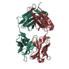
- Assembly
| Deposited unit | 
| ||||||||
|---|---|---|---|---|---|---|---|---|---|
| 1 |
| ||||||||
| Unit cell |
|
-Components
-Antibody , 2 types, 2 molecules HL
| #2: Antibody | Mass: 23800.617 Da / Num. of mol.: 1 / Source method: isolated from a natural source / Source: (natural) Mus musculus (house mouse) |
|---|---|
| #3: Antibody | Mass: 24172.787 Da / Num. of mol.: 1 / Source method: isolated from a natural source / Source: (natural) Mus musculus (house mouse) |
-Protein/peptide / Sugars , 2 types, 2 molecules A
| #1: Protein/peptide | Tau protein / Neurofibrillary tangle protein / Paired helical filament-tau / PHF-tau Mass: 1471.722 Da / Num. of mol.: 1 / Fragment: UNP residues 615-628 / Source method: obtained synthetically / Source: (synth.) Homo sapiens (human) / References: UniProt: P10636 |
|---|---|
| #4: Polysaccharide | alpha-D-glucopyranose-(1-1)-alpha-D-glucopyranose / trehalose / 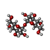  , Oligosaccharide / Class: Nutrient / Mass: 342.297 Da / Num. of mol.: 1 , Oligosaccharide / Class: Nutrient / Mass: 342.297 Da / Num. of mol.: 1Source method: isolated from a genetically manipulated source Details: oligosaccharide with reducing-end-to-reducing-end glycosidic bond References: trehalose |
-Non-polymers , 2 types, 407 molecules 

| #5: Chemical | ChemComp-PGE / Polyethylene glycol Mass: 150.173 Da / Num. of mol.: 4 / Source method: isolated from a natural source / Formula: C6H14O4 Mass: 150.173 Da / Num. of mol.: 4 / Source method: isolated from a natural source / Formula: C6H14O4#6: Water | ChemComp-HOH / | WaterMass: 18.015 Da / Num. of mol.: 403 / Source method: isolated from a natural source / Formula: H2O |
|---|
-Experimental details
-Experiment
| Experiment | Method: X-RAY DIFFRACTION / Number of used crystals: 1 |
|---|
- Sample preparation
| Crystal | Density Matthews: 2.37 Å3/Da / Density % sol: 48.14 % |
|---|---|
| Crystal grow | Temperature: 295 K / Method: vapor diffusion, hanging drop / pH: 8.5 / Details: 30% PEG 4000, 0.1 M Tris, 0.2 M LiSO4 |
-Data collection
| Diffraction | Mean temperature: 100 K | |||||||||||||||||||||||||||||||||||||||||||||||||||||||||||||||||||||||||||||||||||||||||||||||||||||||||||||||||||||||||||||||||||||||||||||||||||
|---|---|---|---|---|---|---|---|---|---|---|---|---|---|---|---|---|---|---|---|---|---|---|---|---|---|---|---|---|---|---|---|---|---|---|---|---|---|---|---|---|---|---|---|---|---|---|---|---|---|---|---|---|---|---|---|---|---|---|---|---|---|---|---|---|---|---|---|---|---|---|---|---|---|---|---|---|---|---|---|---|---|---|---|---|---|---|---|---|---|---|---|---|---|---|---|---|---|---|---|---|---|---|---|---|---|---|---|---|---|---|---|---|---|---|---|---|---|---|---|---|---|---|---|---|---|---|---|---|---|---|---|---|---|---|---|---|---|---|---|---|---|---|---|---|---|---|---|---|
| Diffraction source | Source: SYNCHROTRON / Site: SLS  / Beamline: X06DA / Wavelength: 0.9999 Å / Beamline: X06DA / Wavelength: 0.9999 Å | |||||||||||||||||||||||||||||||||||||||||||||||||||||||||||||||||||||||||||||||||||||||||||||||||||||||||||||||||||||||||||||||||||||||||||||||||||
| Detector | Type: MARMOSAIC 225 mm CCD / Detector: CCD / Date: Dec 5, 2009 | |||||||||||||||||||||||||||||||||||||||||||||||||||||||||||||||||||||||||||||||||||||||||||||||||||||||||||||||||||||||||||||||||||||||||||||||||||
| Radiation | Protocol: SINGLE WAVELENGTH / Monochromatic (M) / Laue (L): M / Scattering type: x-ray | |||||||||||||||||||||||||||||||||||||||||||||||||||||||||||||||||||||||||||||||||||||||||||||||||||||||||||||||||||||||||||||||||||||||||||||||||||
| Radiation wavelength | Wavelength: 0.9999 Å / Relative weight: 1 | |||||||||||||||||||||||||||||||||||||||||||||||||||||||||||||||||||||||||||||||||||||||||||||||||||||||||||||||||||||||||||||||||||||||||||||||||||
| Reflection | Resolution: 1.69→65.44 Å / Num. obs: 41553 / % possible obs: 81.1 % / Observed criterion σ(I): -3 / Redundancy: 3.12 % / Biso Wilson estimate: 34.473 Å2 / CC1/2: 0.999 / Rmerge(I) obs: 0.059 / Rrim(I) all: 0.071 / Χ2: 0.937 / Net I/σ(I): 13.83 / Num. measured all: 129645 / Scaling rejects: 1009 | |||||||||||||||||||||||||||||||||||||||||||||||||||||||||||||||||||||||||||||||||||||||||||||||||||||||||||||||||||||||||||||||||||||||||||||||||||
| Reflection shell |
|
-Phasing
| Phasing | Method: molecular replacement |
|---|
- Processing
| Software |
| ||||||||||||||||||||||||||||||||||||||||||||||||||||||||||||
|---|---|---|---|---|---|---|---|---|---|---|---|---|---|---|---|---|---|---|---|---|---|---|---|---|---|---|---|---|---|---|---|---|---|---|---|---|---|---|---|---|---|---|---|---|---|---|---|---|---|---|---|---|---|---|---|---|---|---|---|---|---|
| Refinement | Method to determine structure: MOLECULAR REPLACEMENT Starting model: 4OZ4 4oz4 Resolution: 1.69→65.44 Å / Cor.coef. Fo:Fc: 0.967 / Cor.coef. Fo:Fc free: 0.95 / SU B: 4.042 / SU ML: 0.119 / Cross valid method: THROUGHOUT / σ(F): 0 / ESU R: 0.142 / ESU R Free: 0.136 Details: HYDROGENS HAVE BEEN ADDED IN THE RIDING POSITIONS U VALUES : REFINED INDIVIDUALLY
| ||||||||||||||||||||||||||||||||||||||||||||||||||||||||||||
| Solvent computation | Ion probe radii: 0.8 Å / Shrinkage radii: 0.8 Å / VDW probe radii: 1.2 Å | ||||||||||||||||||||||||||||||||||||||||||||||||||||||||||||
| Displacement parameters | Biso max: 103.33 Å2 / Biso mean: 29.157 Å2 / Biso min: 15.14 Å2
| ||||||||||||||||||||||||||||||||||||||||||||||||||||||||||||
| Refinement step | Cycle: final / Resolution: 1.69→65.44 Å
| ||||||||||||||||||||||||||||||||||||||||||||||||||||||||||||
| Refine LS restraints |
| ||||||||||||||||||||||||||||||||||||||||||||||||||||||||||||
| LS refinement shell | Resolution: 1.69→1.734 Å / Rfactor Rfree error: 0 / Total num. of bins used: 20
|
