 Movie
Movie Controller


+ Open data

- Basic information
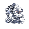
| Entry | Database: PDB / ID: 5mjl | ||||||||||||||||||
|---|---|---|---|---|---|---|---|---|---|---|---|---|---|---|---|---|---|---|---|
| Title | Single-shot pink beam serial crystallography: Proteinase K | ||||||||||||||||||
 Components Components | Proteinase K | ||||||||||||||||||
 Keywords Keywords | HYDROLASE / Proteinase K / Serial Crystallography / Pink beam | ||||||||||||||||||
| Function / homology |  Function and homology information Function and homology informationpeptidase K / serine-type endopeptidase activity / proteolysis / extracellular space / metal ion bindingSimilarity search - Function | ||||||||||||||||||
| Biological species |  Engyodontium album (fungus) Engyodontium album (fungus) | ||||||||||||||||||
| Method | X-RAY DIFFRACTION / SYNCHROTRON / MOLECULAR REPLACEMENT / Resolution: 2.21013789926 Å | ||||||||||||||||||
 Authors Authors | Meents, A. / Oberthuer, D. / Lieske, J. / Srajer, V. | ||||||||||||||||||
| Funding support | 5items
| ||||||||||||||||||
 Citation Citation | Journal: Nat Commun / Year: 2017 Title: Pink-beam serial crystallography. Authors: Meents, A. / Wiedorn, M.O. / Srajer, V. / Henning, R. / Sarrou, I. / Bergtholdt, J. / Barthelmess, M. / Reinke, P.Y.A. / Dierksmeyer, D. / Tolstikova, A. / Schaible, S. / Messerschmidt, M. / ...Authors: Meents, A. / Wiedorn, M.O. / Srajer, V. / Henning, R. / Sarrou, I. / Bergtholdt, J. / Barthelmess, M. / Reinke, P.Y.A. / Dierksmeyer, D. / Tolstikova, A. / Schaible, S. / Messerschmidt, M. / Ogata, C.M. / Kissick, D.J. / Taft, M.H. / Manstein, D.J. / Lieske, J. / Oberthuer, D. / Fischetti, R.F. / Chapman, H.N. #1: Journal: Acta Crystallogr. D Biol. Crystallogr. / Year: 2012 Title: Towards automated crystallographic structure refinement with phenix.refine. Authors: Afonine, P.V. / Grosse-Kunstleve, R.W. / Echols, N. / Headd, J.J. / Moriarty, N.W. / Mustyakimov, M. / Terwilliger, T.C. / Urzhumtsev, A. / Zwart, P.H. / Adams, P.D. #2: Journal: Acta Crystallogr D Biol Crystallogr / Year: 2010 Title: PHENIX: a comprehensive Python-based system for macromolecular structure solution. Authors: Paul D Adams / Pavel V Afonine / Gábor Bunkóczi / Vincent B Chen / Ian W Davis / Nathaniel Echols / Jeffrey J Headd / Li-Wei Hung / Gary J Kapral / Ralf W Grosse-Kunstleve / Airlie J McCoy ...Authors: Paul D Adams / Pavel V Afonine / Gábor Bunkóczi / Vincent B Chen / Ian W Davis / Nathaniel Echols / Jeffrey J Headd / Li-Wei Hung / Gary J Kapral / Ralf W Grosse-Kunstleve / Airlie J McCoy / Nigel W Moriarty / Robert Oeffner / Randy J Read / David C Richardson / Jane S Richardson / Thomas C Terwilliger / Peter H Zwart /  Abstract: Macromolecular X-ray crystallography is routinely applied to understand biological processes at a molecular level. However, significant time and effort are still required to solve and complete many ...Macromolecular X-ray crystallography is routinely applied to understand biological processes at a molecular level. However, significant time and effort are still required to solve and complete many of these structures because of the need for manual interpretation of complex numerical data using many software packages and the repeated use of interactive three-dimensional graphics. PHENIX has been developed to provide a comprehensive system for macromolecular crystallographic structure solution with an emphasis on the automation of all procedures. This has relied on the development of algorithms that minimize or eliminate subjective input, the development of algorithms that automate procedures that are traditionally performed by hand and, finally, the development of a framework that allows a tight integration between the algorithms. | ||||||||||||||||||
| History |
|
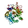
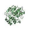
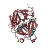
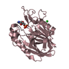
- Structure visualization
| Structure viewer | Molecule: MolmilJmol/JSmol |
|---|
- Downloads & links
-Download
| PDBx/mmCIF format | 5mjl.cif.gz | 143 KB | Display | PDBx/mmCIF format |
|---|---|---|---|---|
| PDB format | pdb5mjl.ent.gz | 89 KB | Display | PDB format |
| PDBx/mmJSON format | 5mjl.json.gz | Tree view | PDBx/mmJSON format | |
| Others |  Other downloads Other downloads |
-Validation report
| Arichive directory | https://data.pdbj.org/pub/pdb/validation_reports/mj/5mjlftp://data.pdbj.org/pub/pdb/validation_reports/mj/5mjl | HTTPS FTP |
|---|
-Related structure data
| Related structure data |  5mjmC  5mjpC 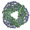 5mjqC  5o7mC 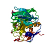 5avjS S: Starting model for refinement C: citing same article ( |
|---|---|
| Similar structure data |


-Links
 PDBj
PDBj

- Assembly
| Deposited unit | 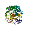
| ||||||||||||
|---|---|---|---|---|---|---|---|---|---|---|---|---|---|
| 1 |
| ||||||||||||
| Unit cell |
|
-Components
-Protein , 1 types, 1 molecules A
| #1: Protein | / Endopeptidase K / Tritirachium alkaline proteinase Mass: 28958.791 Da / Num. of mol.: 1 Source method: isolated from a genetically manipulated source Source: (gene. exp.) Engyodontium album (fungus) / Gene: PROK / Production host: Engyodontium album (fungus) / References: UniProt: P06873, peptidase K |
|---|
-Non-polymers , 5 types, 326 molecules 


| #2: Chemical | ChemComp-CA /  Mass: 40.078 Da / Num. of mol.: 1 / Source method: obtained synthetically / Formula: Ca Mass: 40.078 Da / Num. of mol.: 1 / Source method: obtained synthetically / Formula: Ca |
|---|---|
| #3: Chemical | ChemComp-CL / Chloride Mass: 35.453 Da / Num. of mol.: 1 / Source method: obtained synthetically / Formula: Cl Mass: 35.453 Da / Num. of mol.: 1 / Source method: obtained synthetically / Formula: Cl |
| #4: Chemical | ChemComp-NHE / CHES (buffer) Mass: 207.290 Da / Num. of mol.: 1 / Source method: obtained synthetically / Formula: C8H17NO3S / Comment: pH buffer*YM Mass: 207.290 Da / Num. of mol.: 1 / Source method: obtained synthetically / Formula: C8H17NO3S / Comment: pH buffer*YM |
| #5: Chemical | ChemComp-EPE / HEPES Mass: 238.305 Da / Num. of mol.: 1 / Source method: obtained synthetically / Formula: C8H18N2O4S / Comment: pH buffer*YM Mass: 238.305 Da / Num. of mol.: 1 / Source method: obtained synthetically / Formula: C8H18N2O4S / Comment: pH buffer*YM |
| #6: Water | ChemComp-HOH / WaterMass: 18.015 Da / Num. of mol.: 322 / Source method: isolated from a natural source / Formula: H2O |
-Experimental details
-Experiment
| Experiment | Method: X-RAY DIFFRACTION / Number of used crystals: 1 |
|---|
- Sample preparation
| Crystal | Density Matthews: 2.18 Å3/Da / Density % sol: 43.6 % |
|---|---|
| Crystal grow | Temperature: 293 K / Method: batch mode / Details: 1.6M MgSO4, 10 mM CaCl2, 100 mM CHC buffer pH 6.5 |
-Data collection
| Diffraction | Mean temperature: 293 K | |||||||||
|---|---|---|---|---|---|---|---|---|---|---|
| Diffraction source | Source: SYNCHROTRON / Site: APS / Beamline: 14-BM-D / Wavelength: 1.1-1.3 | |||||||||
| Detector | Type: RAYONIX MX340-HS / Detector: CCD / Date: Jul 10, 2016 | |||||||||
| Radiation | Protocol: LAUE / Monochromatic (M) / Laue (L): L / Scattering type: x-ray | |||||||||
| Radiation wavelength |
| |||||||||
| Reflection | Resolution: 2.1→100 Å / Num. obs: 9197 / % possible obs: 57.9 % / Redundancy: 3.7 % / Biso Wilson estimate: 0.0186601051545 Å2 / Rmerge(I) obs: 0.1 / Net I/σ(I): 18.5 | |||||||||
| Reflection shell | Resolution: 2.21→2.31 Å / Redundancy: 15.31 % / Num. measured obs: 6356 / % possible all: 26.06 |
- Processing
| Software |
| |||||||||||||||||||||||||||||||||||||||||||||||||
|---|---|---|---|---|---|---|---|---|---|---|---|---|---|---|---|---|---|---|---|---|---|---|---|---|---|---|---|---|---|---|---|---|---|---|---|---|---|---|---|---|---|---|---|---|---|---|---|---|---|---|
| Refinement | Method to determine structure: MOLECULAR REPLACEMENT Starting model: 5AVJ Resolution: 2.21013789926→44.1083574029 Å / SU ML: 0.179507088267 / Cross valid method: FREE R-VALUE / σ(F): 0.416332133786 / Phase error: 11.9910233688
| |||||||||||||||||||||||||||||||||||||||||||||||||
| Solvent computation | Shrinkage radii: 0.9 Å / VDW probe radii: 1.11 Å | |||||||||||||||||||||||||||||||||||||||||||||||||
| Displacement parameters | Biso mean: 6.34427693799 Å2 | |||||||||||||||||||||||||||||||||||||||||||||||||
| Refinement step | Cycle: LAST / Resolution: 2.21013789926→44.1083574029 Å
| |||||||||||||||||||||||||||||||||||||||||||||||||
| Refine LS restraints |
| |||||||||||||||||||||||||||||||||||||||||||||||||
| LS refinement shell |
|
