 Movie
Movie Controller

[English] 日本語
 Yorodumi
- PDB-5mdb: The structure of the mature HIV-1 CA hexameric lattice with curva... -
+ Open data

- Basic information
| Entry | Database: PDB / ID: 5mdb | |||||||||
|---|---|---|---|---|---|---|---|---|---|---|
| Title | The structure of the mature HIV-1 CA hexameric lattice with curvature parameters: tilt=17, twist=-6 | |||||||||
 Components Components | (Gag protein HIV-1 protease) x 2 HIV-1 protease) x 2 | |||||||||
 Keywords Keywords | VIRAL PROTEIN / retrovirus / HIV-1 / capsid / lattice curvature | |||||||||
| Function / homology |  Function and homology information Function and homology informationviral budding via host ESCRT complex / viral process / viral capsid / viral nucleocapsid / host cell cytoplasm / virion membrane / structural molecule activity / RNA binding / zinc ion binding / cytoplasmSimilarity search - Function | |||||||||
| Biological species |   Human immunodeficiency virus 1 Human immunodeficiency virus 1 | |||||||||
| Method | ELECTRON MICROSCOPY / subtomogram averaging / cryo EM / Resolution: 8.4 Å | |||||||||
 Authors Authors | Mattei, S. / Glass, B. / Hagen, W.J.H. / Kraeusslich, H.-G. / Briggs, J.A.G. | |||||||||
| Funding support |  Germany, 2items Germany, 2items
| |||||||||
 Citation Citation | Journal: Science / Year: 2016 Title: The structure and flexibility of conical HIV-1 capsids determined within intact virions. Authors: Simone Mattei / Bärbel Glass / Wim J H Hagen / Hans-Georg Kräusslich / John A G Briggs / Abstract: HIV-1 contains a cone-shaped capsid encasing the viral genome. This capsid is thought to follow fullerene geometry-a curved hexameric lattice of the capsid protein, CA, closed by incorporating 12 CA ...HIV-1 contains a cone-shaped capsid encasing the viral genome. This capsid is thought to follow fullerene geometry-a curved hexameric lattice of the capsid protein, CA, closed by incorporating 12 CA pentamers. Current models for core structure are based on crystallography of hexameric and cross-linked pentameric CA, electron microscopy of tubular CA arrays, and simulations. Here, we report subnanometer-resolution cryo-electron tomography structures of hexameric and pentameric CA within intact HIV-1 particles. Whereas the hexamer structure is compatible with crystallography studies, the pentamer forms using different interfaces. Determining multiple structures revealed how CA flexes to form the variably curved core shell. We show that HIV-1 CA assembles both aberrant and perfect fullerene cones, supporting models in which conical cores assemble de novo after maturation. | |||||||||
| History |
|
- Structure visualization
| Movie |
Movie viewer |
|---|---|
| Structure viewer | Molecule: MolmilJmol/JSmol |
- Downloads & links
-Download
| PDBx/mmCIF format | 5mdb.cif.gz | 176 KB | Display | PDBx/mmCIF format |
|---|---|---|---|---|
| PDB format | pdb5mdb.ent.gz | 112.6 KB | Display | PDB format |
| PDBx/mmJSON format | 5mdb.json.gz | Tree view | PDBx/mmJSON format | |
| Others |  Other downloads Other downloads |
-Validation report
| Arichive directory | https://data.pdbj.org/pub/pdb/validation_reports/md/5mdbftp://data.pdbj.org/pub/pdb/validation_reports/md/5mdb | HTTPS FTP |
|---|
-Related structure data
| Related structure data | 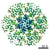 3480MC 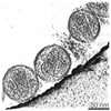 3464C 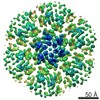 3465C 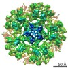 3466C 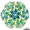 3467C 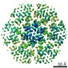 3468C 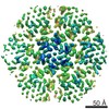 3469C 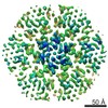 3470C 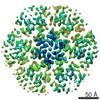 3471C 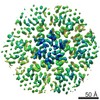 3472C 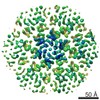 3473C 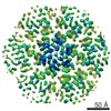 3474C 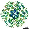 3475C 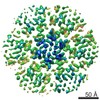 3477C  3478C 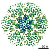 3479C 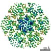 3481C 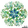 3482C 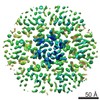 3483C 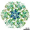 3484C 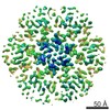 3485C 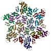 5mcxC 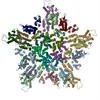 5mcyC 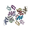 5mczC 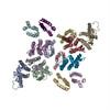 5md0C  5md1C  5md2C  5md3C  5md4C  5md5C  5md6C  5md7C  5md8C  5md9C 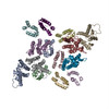 5mdaC 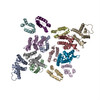 5mdcC 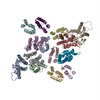 5mddC  5mdeC 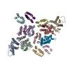 5mdfC  5mdgC M: map data used to model this data C: citing same article ( |
|---|---|
| Similar structure data |
-Links
 PDBj
PDBj

- Assembly
| Deposited unit | 
|
|---|---|
| 1 |
|
-Components
| #1: Protein | HIV-1 protease / Capsid protein p24 C-terminal domain Mass: 8370.568 Da / Num. of mol.: 8 / Source method: isolated from a natural source / Source: (natural) Human immunodeficiency virus 1 / Cell line: MT-4 / Variant: NL43 / References: UniProt: A0A0K0V8J8, UniProt: Q72497*PLUS#2: Protein | HIV-1 protease / Capsid protein p24 N-terminal domainMass: 16301.689 Da / Num. of mol.: 6 / Source method: isolated from a natural source / Source: (natural) Human immunodeficiency virus 1 / Cell line: MT-4 / Variant: NL43 / References: UniProt: E9MJT0, UniProt: Q72497*PLUS |
|---|
-Experimental details
-Experiment
| Experiment | Method: ELECTRON MICROSCOPY |
|---|---|
| EM experiment | Aggregation state: PARTICLE / 3D reconstruction method: subtomogram averaging |
- Sample preparation
| Component | Name: Human immunodeficiency virus 1Subtypes of HIV / Type: VIRUS Details: HIV-1 particles were produced by infection of MT-4 cells with HIV-1 strain NL43 by coculture. Entity ID: all / Source: NATURAL |
|---|---|
| Source (natural) | Organism: Human immunodeficiency virus 1 |
| Details of virus | Empty: NO / Enveloped: YES / Isolate: STRAIN / Type: VIRION |
| Natural host | Organism: Homo sapiens |
| Buffer solution | pH: 7.4 |
| Buffer component | Name: PBS |
| Specimen | Embedding applied: NO / Shadowing applied: NO / Staining applied: NO / Vitrification applied: YES Details: Viral particles were purified from cell culture supernatant by iodixanol gradient centrifugation. |
| Specimen support | Grid material: COPPER / Grid mesh size: 300 divisions/in. / Grid type: C-flat |
| Vitrification | Instrument: FEI VITROBOT MARK II / Cryogen name: ETHANE / Humidity: 100 % / Chamber temperature: 292 K Details: 10nm colloidal gold was added to the sample prior to plunge freezing |
- Electron microscopy imaging
| Experimental equipment |  Model: Titan Krios / Image courtesy: FEI Company |
|---|---|
| Microscopy | Model: FEI TITAN KRIOS / Details: Nanoprobe |
| Electron gun | Electron source: FIELD EMISSION GUN / Accelerating voltage: 300 kV / Illumination mode: FLOOD BEAM |
| Electron lens | Mode: BRIGHT FIELDBright-field microscopy / Nominal magnification: 81000 X / Nominal defocus max: 6500 nm / Nominal defocus min: 2000 nm / Cs: 2.7 mm / C2 aperture diameter: 50 µm / Alignment procedure: ZEMLIN TABLEAU |
| Specimen holder | Cryogen: NITROGEN / Specimen holder model: FEI TITAN KRIOS AUTOGRID HOLDER |
| Image recording | Electron dose: 2.2 e/Å2 / Detector mode: SUPER-RESOLUTION / Film or detector model: GATAN K2 QUANTUM (4k x 4k) / Num. of grids imaged: 1 |
| EM imaging optics | Energyfilter name: GIF Quantum LS / Energyfilter upper: 20 eV / Energyfilter lower: 0 eV |
| Image scans | Width: 3710 / Height: 3838 / Movie frames/image: 5 |
- Processing
| EM software |
| ||||||||||||||||||||||||
|---|---|---|---|---|---|---|---|---|---|---|---|---|---|---|---|---|---|---|---|---|---|---|---|---|---|
| Image processing | Details: Frames were aligned using MotionCorr. Tilts in a tilt series were exposure filtered for cumulative electron dose. Tomograms were reconstructed using IMOD. | ||||||||||||||||||||||||
| CTF correction | Details: CTF correction was performed using the ctfphaseflip program in IMOD prior to backprojection. Type: PHASE FLIPPING ONLY | ||||||||||||||||||||||||
| Symmetry | Point symmetry: C1 (asymmetric) | ||||||||||||||||||||||||
| 3D reconstruction | Resolution: 8.4 Å / Resolution method: FSC 0.143 CUT-OFF / Num. of particles: 17092 Details: The final reconstruction is obtained by averaging, without further alignment, subtomograms that were selected based on the local curvature of the hexameric lattice. This reconstruction has ...Details: The final reconstruction is obtained by averaging, without further alignment, subtomograms that were selected based on the local curvature of the hexameric lattice. This reconstruction has hexamer-hexamer curvature parameters: tilt=17, twist=-6. Num. of class averages: 1 / Symmetry type: POINT | ||||||||||||||||||||||||
| EM volume selection | Details: Subtomograms were extracted along the manually rendered core surface of each viral particle. Num. of tomograms: 103 / Num. of volumes extracted: 652618 / Reference model: reference free | ||||||||||||||||||||||||
| Atomic model building | Protocol: RIGID BODY FIT / Space: REAL / Target criteria: Cross-correlation coefficient |
