 Movie
Movie Controller

[English] 日本語
 Yorodumi
- PDB-5mb4: Crystal Structure of the Psathyrella asperospora lectin PAL in co... -
+ Open data

- Basic information
| Entry | Database: PDB / ID: 5mb4 | |||||||||
|---|---|---|---|---|---|---|---|---|---|---|
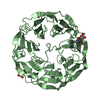
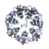
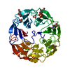
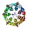
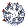
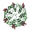
| Title | Crystal Structure of the Psathyrella asperospora lectin PAL in complex with GlcNAc | |||||||||
 Components Components | GlcNAc specific lectin | |||||||||
 Keywords Keywords | SUGAR BINDING PROTEIN /  Lectin / fungi / seven bladed beta propeller / GlcNAc Lectin / fungi / seven bladed beta propeller / GlcNAc | |||||||||
| Function / homology | FG-GAP-like repeat / FG-GAP repeat / MALONATE ION / 2-acetamido-2-deoxy-alpha-D-glucopyranose / GlcNAc specific lectin Function and homology information Function and homology information | |||||||||
| Biological species |  Psathyrella (fungus) Psathyrella (fungus) | |||||||||
| Method | X-RAY DIFFRACTION / SYNCHROTRON / MOLECULAR REPLACEMENT / Resolution: 2 Å | |||||||||
 Authors Authors | Ribeiro, J. / Varrot, A. | |||||||||
| Funding support |  France, 1items France, 1items
| |||||||||
 Citation Citation | Journal: Proteins / Year: 2017 Title: Biophysical characterization and structural determination of the potent cytotoxic Psathyrella asperospora lectin. Authors: Ribeiro, J.P. / Ali Abol Hassan, M. / Rouf, R. / Tiralongo, E. / May, T.W. / Day, C.J. / Imberty, A. / Tiralongo, J. / Varrot, A. | |||||||||
| History |
|
- Structure visualization
| Structure viewer | Molecule: MolmilJmol/JSmol |
|---|
- Downloads & links
-Download
| PDBx/mmCIF format | 5mb4.cif.gz | 102.1 KB | Display | PDBx/mmCIF format |
|---|---|---|---|---|
| PDB format | pdb5mb4.ent.gz | 77.2 KB | Display | PDB format |
| PDBx/mmJSON format | 5mb4.json.gz | Tree view | PDBx/mmJSON format | |
| Others |  Other downloads Other downloads |
-Validation report
| Arichive directory | https://data.pdbj.org/pub/pdb/validation_reports/mb/5mb4ftp://data.pdbj.org/pub/pdb/validation_reports/mb/5mb4 | HTTPS FTP |
|---|
-Related structure data
| Related structure data | 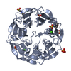 2bwrS S: Starting model for refinement |
|---|---|
| Similar structure data |


-Links
 PDBj
PDBj


- Assembly
| Deposited unit | 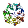
| ||||||||||||
|---|---|---|---|---|---|---|---|---|---|---|---|---|---|
| 1 |
| ||||||||||||
| Unit cell |
| ||||||||||||
| Components on special symmetry positions |
|
-Components
-Protein , 1 types, 1 molecules A
| #1: Protein | Mass: 42706.629 Da / Num. of mol.: 1 / Source method: isolated from a natural source / Source: (natural) Psathyrella (fungus) / Variant: Asperospora / References: UniProt: A0A1U7Q1Z0*PLUS |
|---|
-Sugars , 2 types, 8 molecules 

| #4: Sugar | ChemComp-NAG / N-Acetylglucosamine Type: D-saccharide, beta linking / Mass: 221.208 Da / Num. of mol.: 6 Type: D-saccharide, beta linking / Mass: 221.208 Da / Num. of mol.: 6Source method: isolated from a genetically manipulated source Formula: C8H15NO6 #5: Sugar | N-Acetylglucosamine Type: D-saccharide, alpha linking / Mass: 221.208 Da / Num. of mol.: 2 Type: D-saccharide, alpha linking / Mass: 221.208 Da / Num. of mol.: 2Source method: isolated from a genetically manipulated source Formula: C8H15NO6 |
|---|
-Non-polymers , 3 types, 354 molecules 


| #2: Chemical | ChemComp-NA /  Mass: 22.990 Da / Num. of mol.: 6 / Source method: obtained synthetically / Formula: Na Mass: 22.990 Da / Num. of mol.: 6 / Source method: obtained synthetically / Formula: Na#3: Chemical | ChemComp-MLI / | Malonic acid Mass: 102.046 Da / Num. of mol.: 1 / Source method: obtained synthetically / Formula: C3H2O4 Mass: 102.046 Da / Num. of mol.: 1 / Source method: obtained synthetically / Formula: C3H2O4#6: Water | ChemComp-HOH / | WaterMass: 18.015 Da / Num. of mol.: 347 / Source method: isolated from a natural source / Formula: H2O |
|---|
-Experimental details
-Experiment
| Experiment | Method: X-RAY DIFFRACTION / Number of used crystals: 1 |
|---|
- Sample preparation
| Crystal | Density Matthews: 3.27 Å3/Da / Density % sol: 62.35 % / Description: plate |
|---|---|
| Crystal grow | Temperature: 292 K / Method: vapor diffusion, hanging drop / pH: 5 Details: Protein at 10 mg per ml in PBS 2.4M sodium malonate pH 5.0 |
-Data collection
| Diffraction | Mean temperature: 100 K |
|---|---|
| Diffraction source | Source: SYNCHROTRON / Site: ESRF / Beamline: ID29 / Wavelength: 0.979 Å |
| Detector | Type: DECTRIS PILATUS 6M / Detector: PIXEL / Date: Oct 5, 2014 |
| Radiation | Protocol: SINGLE WAVELENGTH / Monochromatic (M) / Laue (L): M / Scattering type: x-ray |
| Radiation wavelength | Wavelength: 0.979 Å / Relative weight: 1 |
| Reflection | Resolution: 2→30.91 Å / Num. obs: 37041 / % possible obs: 98.5 % / Observed criterion σ(I): 2 / Redundancy: 3.1 % / Biso Wilson estimate: 26.3 Å2 / CC1/2: 0.996 / Rmerge(I) obs: 0.078 / Net I/av σ(I): 7.5 / Net I/σ(I): 7.8 |
| Reflection shell | Resolution: 2→2.05 Å / Redundancy: 3 % / Rmerge(I) obs: 0.41 / Mean I/σ(I) obs: 2.2 / CC1/2: 0.608 / % possible all: 99.4 |
- Processing
| Software |
| ||||||||||||||||||||||||||||||||||||||||||||||||||||||||||||||||||||||||||||||||||||||||||||||||||||||||||||||||||||||||||||||||||||||||||||||||||||||||||||||||||||||||||||||||||||||
|---|---|---|---|---|---|---|---|---|---|---|---|---|---|---|---|---|---|---|---|---|---|---|---|---|---|---|---|---|---|---|---|---|---|---|---|---|---|---|---|---|---|---|---|---|---|---|---|---|---|---|---|---|---|---|---|---|---|---|---|---|---|---|---|---|---|---|---|---|---|---|---|---|---|---|---|---|---|---|---|---|---|---|---|---|---|---|---|---|---|---|---|---|---|---|---|---|---|---|---|---|---|---|---|---|---|---|---|---|---|---|---|---|---|---|---|---|---|---|---|---|---|---|---|---|---|---|---|---|---|---|---|---|---|---|---|---|---|---|---|---|---|---|---|---|---|---|---|---|---|---|---|---|---|---|---|---|---|---|---|---|---|---|---|---|---|---|---|---|---|---|---|---|---|---|---|---|---|---|---|---|---|---|---|
| Refinement | Method to determine structure: MOLECULAR REPLACEMENT Starting model: 2bwr Resolution: 2→30.91 Å / Cor.coef. Fo:Fc: 0.968 / Cor.coef. Fo:Fc free: 0.946 / SU B: 4.643 / SU ML: 0.12 / Cross valid method: THROUGHOUT / ESU R: 0.142 / ESU R Free: 0.139 / Details: HYDROGENS HAVE BEEN ADDED IN THE RIDING POSITIONS
| ||||||||||||||||||||||||||||||||||||||||||||||||||||||||||||||||||||||||||||||||||||||||||||||||||||||||||||||||||||||||||||||||||||||||||||||||||||||||||||||||||||||||||||||||||||||
| Solvent computation | Ion probe radii: 0.8 Å / Shrinkage radii: 0.8 Å / VDW probe radii: 1.2 Å | ||||||||||||||||||||||||||||||||||||||||||||||||||||||||||||||||||||||||||||||||||||||||||||||||||||||||||||||||||||||||||||||||||||||||||||||||||||||||||||||||||||||||||||||||||||||
| Displacement parameters | Biso mean: 28.193 Å2
| ||||||||||||||||||||||||||||||||||||||||||||||||||||||||||||||||||||||||||||||||||||||||||||||||||||||||||||||||||||||||||||||||||||||||||||||||||||||||||||||||||||||||||||||||||||||
| Refinement step | Cycle: 1 / Resolution: 2→30.91 Å
| ||||||||||||||||||||||||||||||||||||||||||||||||||||||||||||||||||||||||||||||||||||||||||||||||||||||||||||||||||||||||||||||||||||||||||||||||||||||||||||||||||||||||||||||||||||||
| Refine LS restraints |
|