 Movie
Movie Controller


+ Open data

- Basic information
| Entry | Database: PDB / ID: 5lsv | |||||||||
|---|---|---|---|---|---|---|---|---|---|---|
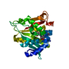
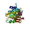
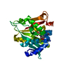
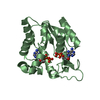
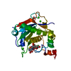
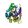
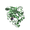
| Title | X-ray crystal structure of AA13 LPMO | |||||||||
 Components Components | AoAA13 | |||||||||
 Keywords Keywords | METAL BINDING PROTEIN /  ENZYME / ASPERGILLUS ORYZAE AA13 LPMO ENZYME / ASPERGILLUS ORYZAE AA13 LPMO | |||||||||
| Function / homology | Cellulose/chitin-binding protein, N-terminal / Lytic polysaccharide mono-oxygenase, cellulose-degrading / membrane / metal ion binding / alpha-maltose / Inactive lytic polysaccharide monooxygenase Function and homology information Function and homology information | |||||||||
| Biological species |  Aspergillus oryzae RIB40 (mold) Aspergillus oryzae RIB40 (mold) | |||||||||
| Method | X-RAY DIFFRACTION / SYNCHROTRON / MOLECULAR REPLACEMENT / Resolution: 1.1 Å | |||||||||
 Authors Authors | Frandsen, K.E.H. / Poulsen, J.-C.N. / Tovborg, M. / Johansen, K.S. / Lo Leggio, L. | |||||||||
| Funding support |  Denmark, 1items Denmark, 1items
| |||||||||
 Citation Citation | Journal: Acta Crystallogr D Struct Biol / Year: 2017 Title: Learning from oligosaccharide soaks of crystals of an AA13 lytic polysaccharide monooxygenase: crystal packing, ligand binding and active-site disorder. Authors: Frandsen, K.E. / Poulsen, J.C. / Tovborg, M. / Johansen, K.S. / Lo Leggio, L. | |||||||||
| History |
|
- Structure visualization
| Structure viewer | Molecule: MolmilJmol/JSmol |
|---|
- Downloads & links
-Download
| PDBx/mmCIF format | 5lsv.cif.gz | 136.1 KB | Display | PDBx/mmCIF format |
|---|---|---|---|---|
| PDB format | pdb5lsv.ent.gz | 105.1 KB | Display | PDB format |
| PDBx/mmJSON format | 5lsv.json.gz | Tree view | PDBx/mmJSON format | |
| Others |  Other downloads Other downloads |
-Validation report
| Arichive directory | https://data.pdbj.org/pub/pdb/validation_reports/ls/5lsvftp://data.pdbj.org/pub/pdb/validation_reports/ls/5lsv | HTTPS FTP |
|---|
-Related structure data
| Related structure data |  5t7jC  5t7kC  5t7nC 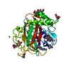 4opbS S: Starting model for refinement C: citing same article ( |
|---|---|
| Similar structure data |


-Links
 PDBj
PDBj


- Assembly
| Deposited unit | 
| ||||||||
|---|---|---|---|---|---|---|---|---|---|
| 1 |
| ||||||||
| Unit cell |
|
-Components
| #1: Protein | Mass: 25691.057 Da / Num. of mol.: 1 Source method: isolated from a genetically manipulated source Source: (gene. exp.) Aspergillus oryzae RIB40 (mold) / Gene: AO090701000246 / Production host: Aspergillus oryzae RIB40 (mold) / References: UniProt: Q2U8Y3 | ||
|---|---|---|---|
| #2: Polysaccharide | alpha-D-glucopyranose-(1-4)-alpha-D-glucopyranose / alpha-maltose  , Oligosaccharide / Class: Nutrient / Mass: 342.297 Da / Num. of mol.: 1 , Oligosaccharide / Class: Nutrient / Mass: 342.297 Da / Num. of mol.: 1Source method: isolated from a genetically manipulated source Details: oligosaccharide / References: alpha-maltose | ||
| #3: Sugar | ChemComp-NAG / N-Acetylglucosamine  Type: D-saccharide, beta linking / Mass: 221.208 Da / Num. of mol.: 1 Type: D-saccharide, beta linking / Mass: 221.208 Da / Num. of mol.: 1Source method: isolated from a genetically manipulated source Formula: C8H15NO6 | ||
| #4: Chemical | ChemComp-ZN /   Mass: 65.409 Da / Num. of mol.: 8 / Source method: isolated from a natural source / Formula: Zn Mass: 65.409 Da / Num. of mol.: 8 / Source method: isolated from a natural source / Formula: Zn#5: Water | ChemComp-HOH / | Water Mass: 18.015 Da / Num. of mol.: 354 / Source method: isolated from a natural source / Formula: H2O Mass: 18.015 Da / Num. of mol.: 354 / Source method: isolated from a natural source / Formula: H2O |
-Experimental details
-Experiment
| Experiment | Method: X-RAY DIFFRACTION / Number of used crystals: 1 |
|---|
- Sample preparation
| Crystal | Density Matthews: 1.87 Å3/Da / Density % sol: 34.19 % |
|---|---|
| Crystal grow | Temperature: 298 K / Method: vapor diffusion, hanging drop / pH: 5 Details: 20%(v/v)PEG3000 0.2M Zn-acetate 0.1 M Malate/MES/Tris pH5.0 PH range: 4.5-8.0 |
-Data collection
| Diffraction | Mean temperature: 100 K |
|---|---|
| Diffraction source | Source: SYNCHROTRON / Site: MAX II  / Beamline: I911-3 / Wavelength: 1 Å / Beamline: I911-3 / Wavelength: 1 Å |
| Detector | Type: MAR CCD 165 mm / Detector: CCD / Date: May 21, 2013 |
| Radiation | Monochromator: SI(111) / Protocol: SINGLE WAVELENGTH / Monochromatic (M) / Laue (L): M / Scattering type: x-ray |
| Radiation wavelength | Wavelength: 1 Å / Relative weight: 1 |
| Reflection | Resolution: 1.1→30 Å / Num. obs: 78799 / % possible obs: 99.9 % / Redundancy: 7.44 % / Rrim(I) all: 0.129 / Net I/σ(I): 13.37 |
| Reflection shell | Resolution: 1.1→1.13 Å / Redundancy: 6.76 % / Rrim(I) all: 0.9 / % possible all: 99.9 |
- Processing
| Software |
| ||||||||||||||||||||||||||||||||||||||||||||||||||||||||||||||||||||||||||||||||||||||||||||||||||||||||||||||||||||||||||||||||||||||||||||||||||||||||||||||||||||||||||||||||||||||
|---|---|---|---|---|---|---|---|---|---|---|---|---|---|---|---|---|---|---|---|---|---|---|---|---|---|---|---|---|---|---|---|---|---|---|---|---|---|---|---|---|---|---|---|---|---|---|---|---|---|---|---|---|---|---|---|---|---|---|---|---|---|---|---|---|---|---|---|---|---|---|---|---|---|---|---|---|---|---|---|---|---|---|---|---|---|---|---|---|---|---|---|---|---|---|---|---|---|---|---|---|---|---|---|---|---|---|---|---|---|---|---|---|---|---|---|---|---|---|---|---|---|---|---|---|---|---|---|---|---|---|---|---|---|---|---|---|---|---|---|---|---|---|---|---|---|---|---|---|---|---|---|---|---|---|---|---|---|---|---|---|---|---|---|---|---|---|---|---|---|---|---|---|---|---|---|---|---|---|---|---|---|---|---|
| Refinement | Method to determine structure: MOLECULAR REPLACEMENT Starting model: 4OPB Resolution: 1.1→30 Å / Cor.coef. Fo:Fc: 0.986 / Cor.coef. Fo:Fc free: 0.981 / SU B: 0.837 / SU ML: 0.018 / Cross valid method: THROUGHOUT / ESU R: 0.024 / ESU R Free: 0.025 / Details: HYDROGENS HAVE BEEN ADDED IN THE RIDING POSITIONS
| ||||||||||||||||||||||||||||||||||||||||||||||||||||||||||||||||||||||||||||||||||||||||||||||||||||||||||||||||||||||||||||||||||||||||||||||||||||||||||||||||||||||||||||||||||||||
| Solvent computation | Ion probe radii: 0.8 Å / Shrinkage radii: 0.8 Å / VDW probe radii: 1.2 Å | ||||||||||||||||||||||||||||||||||||||||||||||||||||||||||||||||||||||||||||||||||||||||||||||||||||||||||||||||||||||||||||||||||||||||||||||||||||||||||||||||||||||||||||||||||||||
| Displacement parameters | Biso mean: 8.816 Å2
| ||||||||||||||||||||||||||||||||||||||||||||||||||||||||||||||||||||||||||||||||||||||||||||||||||||||||||||||||||||||||||||||||||||||||||||||||||||||||||||||||||||||||||||||||||||||
| Refinement step | Cycle: LAST / Resolution: 1.1→30 Å
| ||||||||||||||||||||||||||||||||||||||||||||||||||||||||||||||||||||||||||||||||||||||||||||||||||||||||||||||||||||||||||||||||||||||||||||||||||||||||||||||||||||||||||||||||||||||
| Refine LS restraints |
|