 Movie
Movie Controller


+ Open data

- Basic information
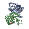
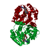
| Entry | Database: PDB / ID: 5log | ||||||
|---|---|---|---|---|---|---|---|
| Title | Crystal Structure of SafC from Myxococcus xanthus bound to SAM | ||||||
 Components Components | Putative O-methyltransferase | ||||||
 Keywords Keywords | TRANSFERASE / O-METHYL TRANSFERASE / SAM | ||||||
| Function / homology |  Function and homology information Function and homology information | ||||||
| Biological species |  Myxococcus xanthus (bacteria) Myxococcus xanthus (bacteria) | ||||||
| Method | X-RAY DIFFRACTION / SYNCHROTRON / MOLECULAR REPLACEMENT / Resolution: 2.01 Å | ||||||
 Authors Authors | Gerhardt, S. / Netzer, J. / Einsle, O. | ||||||
 Citation Citation | Journal: FEBS Lett. / Year: 2017 Title: Functional and structural characterisation of a bacterial O-methyltransferase and factors determining regioselectivity. Authors: Siegrist, J. / Netzer, J. / Mordhorst, S. / Karst, L. / Gerhardt, S. / Einsle, O. / Richter, M. / Andexer, J.N. | ||||||
| History |
|
- Structure visualization
| Structure viewer | Molecule: MolmilJmol/JSmol |
|---|
- Downloads & links
-Download
| PDBx/mmCIF format | 5log.cif.gz | 195.1 KB | Display | PDBx/mmCIF format |
|---|---|---|---|---|
| PDB format | pdb5log.ent.gz | 152.5 KB | Display | PDB format |
| PDBx/mmJSON format | 5log.json.gz | Tree view | PDBx/mmJSON format | |
| Others |  Other downloads Other downloads |
-Validation report
| Arichive directory | https://data.pdbj.org/pub/pdb/validation_reports/lo/5logftp://data.pdbj.org/pub/pdb/validation_reports/lo/5log | HTTPS FTP |
|---|
-Related structure data
| Related structure data |  5lhmC  2hnkS S: Starting model for refinement C: citing same article ( |
|---|---|
| Similar structure data |


-Links
 PDBj
PDBj- Assembly
| Deposited unit | 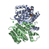
| ||||||||
|---|---|---|---|---|---|---|---|---|---|
| 1 |
| ||||||||
| Unit cell |
|
-Components
-Protein , 1 types, 2 molecules AB
| #1: Protein | Mass: 26262.963 Da / Num. of mol.: 2 / Mutation: T78A Source method: isolated from a genetically manipulated source Source: (gene. exp.) Myxococcus xanthus (bacteria) / Gene: safC / Plasmid: pET28A / Production host: Escherichia coli (E. coli) / Strain (production host): BL21(DE3) / References: UniProt: Q50859 |
|---|
-Non-polymers , 5 types, 302 molecules 


| #2: Chemical | ChemComp-SAH / S-Adenosyl-L-homocysteine Type: L-peptide linking / Mass: 384.411 Da / Num. of mol.: 1 / Source method: obtained synthetically / Formula: C14H20N6O5S Type: L-peptide linking / Mass: 384.411 Da / Num. of mol.: 1 / Source method: obtained synthetically / Formula: C14H20N6O5S | ||||||
|---|---|---|---|---|---|---|---|
| #3: Chemical |  Mass: 24.305 Da / Num. of mol.: 2 / Source method: obtained synthetically / Formula: Mg Mass: 24.305 Da / Num. of mol.: 2 / Source method: obtained synthetically / Formula: Mg#4: Chemical | ChemComp-CL / | Chloride Mass: 35.453 Da / Num. of mol.: 1 / Source method: obtained synthetically / Formula: Cl Mass: 35.453 Da / Num. of mol.: 1 / Source method: obtained synthetically / Formula: Cl#5: Chemical | ChemComp-LDP / | Dopamine (medication) Mass: 153.178 Da / Num. of mol.: 1 / Source method: obtained synthetically / Formula: C8H11NO2 / Comment: medication*YM Mass: 153.178 Da / Num. of mol.: 1 / Source method: obtained synthetically / Formula: C8H11NO2 / Comment: medication*YM#6: Water | ChemComp-HOH / | WaterMass: 18.015 Da / Num. of mol.: 297 / Source method: isolated from a natural source / Formula: H2O |
-Experimental details
-Experiment
| Experiment | Method: X-RAY DIFFRACTION / Number of used crystals: 1 |
|---|
- Sample preparation
| Crystal | Density Matthews: 2.13 Å3/Da / Density % sol: 42.14 % / Mosaicity: 0.12 ° |
|---|---|
| Crystal grow | Temperature: 298 K / Method: vapor diffusion, sitting drop / pH: 5.5 / Details: PEG MME 5000, Sodium Acetate pH 5.5 |
-Data collection
| Diffraction | Mean temperature: 100 K |
|---|---|
| Diffraction source | Source: SYNCHROTRON / Site: SLS  / Beamline: X06DA / Wavelength: 1.00003 Å / Beamline: X06DA / Wavelength: 1.00003 Å |
| Detector | Type: DECTRIS PILATUS3 S 6M / Detector: PIXEL / Date: Aug 24, 2014 / Details: mirrors |
| Radiation | Monochromator: Si(111) / Protocol: SINGLE WAVELENGTH / Monochromatic (M) / Laue (L): M / Scattering type: x-ray |
| Radiation wavelength | Wavelength: 1.00003 Å / Relative weight: 1 |
| Reflection | Resolution: 2.005→71.86 Å / Num. obs: 28956 / % possible obs: 97 % / Redundancy: 6.7 % / Biso Wilson estimate: 19.58 Å2 / CC1/2: 0.98 / Rmerge(I) obs: 0.303 / Rpim(I) all: 0.137 / Rrim(I) all: 0.357 / Rsym value: 0.359 / Net I/σ(I): 7 / Num. measured all: 193880 / Scaling rejects: 82 |
| Reflection shell | Resolution: 2.005→2.11 Å / Redundancy: 6.8 % / Rmerge(I) obs: 1.159 / Mean I/σ(I) obs: 2.1 / CC1/2: 0.373 / % possible all: 90.4 |
- Processing
| Software |
| ||||||||||||||||||||||||||||||||||||||||||||||||||||||||||||||||||||||||||||||||||||||||||||||||||||||||||||
|---|---|---|---|---|---|---|---|---|---|---|---|---|---|---|---|---|---|---|---|---|---|---|---|---|---|---|---|---|---|---|---|---|---|---|---|---|---|---|---|---|---|---|---|---|---|---|---|---|---|---|---|---|---|---|---|---|---|---|---|---|---|---|---|---|---|---|---|---|---|---|---|---|---|---|---|---|---|---|---|---|---|---|---|---|---|---|---|---|---|---|---|---|---|---|---|---|---|---|---|---|---|---|---|---|---|---|---|---|---|
| Refinement | Method to determine structure: MOLECULAR REPLACEMENT Starting model: 2HNK Resolution: 2.01→71.86 Å / Cor.coef. Fo:Fc: 0.934 / Cor.coef. Fo:Fc free: 0.919 / Rfactor Rfree error: 0 / SU R Cruickshank DPI: 0.189 / Cross valid method: THROUGHOUT / σ(F): 0 / SU R Blow DPI: 0.197 / SU Rfree Blow DPI: 0.158 / SU Rfree Cruickshank DPI: 0.156
| ||||||||||||||||||||||||||||||||||||||||||||||||||||||||||||||||||||||||||||||||||||||||||||||||||||||||||||
| Displacement parameters | Biso max: 110.64 Å2 / Biso mean: 27.71 Å2 / Biso min: 4.89 Å2
| ||||||||||||||||||||||||||||||||||||||||||||||||||||||||||||||||||||||||||||||||||||||||||||||||||||||||||||
| Refine analyze | Luzzati coordinate error obs: 0.25 Å | ||||||||||||||||||||||||||||||||||||||||||||||||||||||||||||||||||||||||||||||||||||||||||||||||||||||||||||
| Refinement step | Cycle: final / Resolution: 2.01→71.86 Å
| ||||||||||||||||||||||||||||||||||||||||||||||||||||||||||||||||||||||||||||||||||||||||||||||||||||||||||||
| Refine LS restraints |
| ||||||||||||||||||||||||||||||||||||||||||||||||||||||||||||||||||||||||||||||||||||||||||||||||||||||||||||
| LS refinement shell | Resolution: 2.01→2.09 Å / Total num. of bins used: 14
| ||||||||||||||||||||||||||||||||||||||||||||||||||||||||||||||||||||||||||||||||||||||||||||||||||||||||||||
| Refinement TLS params. | Method: refined / Refine-ID: X-RAY DIFFRACTION
| ||||||||||||||||||||||||||||||||||||||||||||||||||||||||||||||||||||||||||||||||||||||||||||||||||||||||||||
| Refinement TLS group |
|
