 Movie
Movie Controller

[English] 日本語
 Yorodumi
- PDB-5lkh: Cryo-EM structure of the Tc toxin TcdA1 in its pore state (obtain... -
+ Open data

- Basic information
| Entry | Database: PDB / ID: 5lkh | ||||||
|---|---|---|---|---|---|---|---|
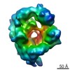
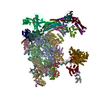
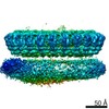
| Title | Cryo-EM structure of the Tc toxin TcdA1 in its pore state (obtained by flexible fitting) | ||||||
 Components Components | TcdA1 | ||||||
 Keywords Keywords |  TOXIN / nanodisc / injection / pore-forming TOXIN / nanodisc / injection / pore-forming | ||||||
| Function / homology |  Function and homology information Function and homology information | ||||||
| Biological species |  Photorhabdus luminescens (bacteria) Photorhabdus luminescens (bacteria) | ||||||
| Method | ELECTRON MICROSCOPY / single particle reconstruction / cryo EM / Resolution: 3.46 Å | ||||||
 Authors Authors | Gatsogiannis, C. / Merino, F. / Prumbaum, D. / Roderer, D. / Leidreiter, F. / Meusch, D. / Raunser, S. | ||||||
 Citation Citation | Journal: Nat Struct Mol Biol / Year: 2016 Title: Membrane insertion of a Tc toxin in near-atomic detail. Authors: Christos Gatsogiannis / Felipe Merino / Daniel Prumbaum / Daniel Roderer / Franziska Leidreiter / Dominic Meusch / Stefan Raunser /  Abstract: Tc toxins from pathogenic bacteria use a special syringe-like mechanism to perforate the host cell membrane and inject a deadly enzyme into the host cytosol. The molecular mechanism of this unusual ...Tc toxins from pathogenic bacteria use a special syringe-like mechanism to perforate the host cell membrane and inject a deadly enzyme into the host cytosol. The molecular mechanism of this unusual injection system is poorly understood. Using electron cryomicroscopy, we determined the structure of TcdA1 from Photorhabdus luminescens embedded in lipid nanodiscs. In our structure, compared with the previous structure of TcdA1 in the prepore state, the transmembrane helices rearrange in the membrane and open the initially closed pore. However, the helices do not span the complete membrane; instead, the loops connecting the helices form the rim of the funnel. Lipid head groups reach into the space between the loops and consequently stabilize the pore conformation. The linker domain is folded and packed into a pocket formed by the other domains of the toxin, thereby considerably contributing to stabilization of the pore state. | ||||||
| History |
|
- Structure visualization
| Movie |
Movie viewer |
|---|---|
| Structure viewer | Molecule: MolmilJmol/JSmol |
- Downloads & links
-Download
| PDBx/mmCIF format | 5lkh.cif.gz | 925.9 KB | Display | PDBx/mmCIF format |
|---|---|---|---|---|
| PDB format | pdb5lkh.ent.gz | 719.6 KB | Display | PDB format |
| PDBx/mmJSON format | 5lkh.json.gz | Tree view | PDBx/mmJSON format | |
| Others |  Other downloads Other downloads |
-Validation report
| Arichive directory | https://data.pdbj.org/pub/pdb/validation_reports/lk/5lkhftp://data.pdbj.org/pub/pdb/validation_reports/lk/5lkh | HTTPS FTP |
|---|
-Related structure data
| Related structure data | 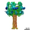 4068MC  5lkiC M: map data used to model this data C: citing same article ( |
|---|---|
| Similar structure data |
-Links
 PDBj
PDBj
- Assembly
| Deposited unit | 
|
|---|---|
| 1 |
|
-Components
| #1: Protein | Mass: 283229.406 Da / Num. of mol.: 5 Source method: isolated from a genetically manipulated source Source: (gene. exp.) Photorhabdus luminescens (bacteria) / Gene: tcdA, tcdA1 / Production host: Escherichia coli (E. coli) / References: UniProt: Q9RN43 |
|---|
-Experimental details
-Experiment
| Experiment | Method: ELECTRON MICROSCOPY |
|---|---|
| EM experiment | Aggregation state: PARTICLE / 3D reconstruction method: single particle reconstruction |
- Sample preparation
| Component | Name: Structure of TcdA1 in pore state, determined in lipid nanodiscs Type: COMPLEX / Entity ID: all / Source: RECOMBINANT | ||||||||||||
|---|---|---|---|---|---|---|---|---|---|---|---|---|---|
| Molecular weight | Value: 1.4 MDa / Experimental value: NO | ||||||||||||
| Source (natural) | Organism: Photorhabdus luminescens (bacteria) | ||||||||||||
| Source (recombinant) | Organism: Escherichia coli (E. coli) / Plasmid: pET-19b | ||||||||||||
| Buffer solution | pH: 11 | ||||||||||||
| Buffer component |
| ||||||||||||
| Specimen | Embedding applied: NO / Shadowing applied: NO / Staining applied: NO / Vitrification applied: YES | ||||||||||||
| Specimen support | Grid material: COPPER / Grid mesh size: 400 divisions/in. / Grid type: C-flat-2/1 | ||||||||||||
| Vitrification | Instrument: GATAN CRYOPLUNGE 3 / Cryogen name: ETHANE / Humidity: 90 % |
- Electron microscopy imaging
| Experimental equipment |  Model: Titan Krios / Image courtesy: FEI Company |
|---|---|
| Microscopy | Model: FEI TITAN KRIOS / Details: Cs corrected microscope |
| Electron gun | Electron source: FIELD EMISSION GUN / Accelerating voltage: 300 kV / Illumination mode: OTHER |
| Electron lens | Mode: BRIGHT FIELDBright-field microscopy / Nominal magnification: 59000 X / Alignment procedure: COMA FREE |
| Specimen holder | Cryogen: NITROGEN |
| Image recording | Electron dose: 15.4 e/Å2 / Film or detector model: FEI FALCON II (4k x 4k) / Num. of real images: 1957 |
| Image scans | Movie frames/image: 7 |
- Processing
| EM software |
| ||||||||||||||||||||||||||||||||
|---|---|---|---|---|---|---|---|---|---|---|---|---|---|---|---|---|---|---|---|---|---|---|---|---|---|---|---|---|---|---|---|---|---|
| CTF correction | Type: PHASE FLIPPING AND AMPLITUDE CORRECTION | ||||||||||||||||||||||||||||||||
| Particle selection | Num. of particles selected: 30061 / Details: particles were picked manually | ||||||||||||||||||||||||||||||||
| Symmetry | Point symmetry: C5 (5 fold cyclic) | ||||||||||||||||||||||||||||||||
| 3D reconstruction | Resolution: 3.46 Å / Resolution method: FSC 0.143 CUT-OFF / Num. of particles: 13000 Details: The density was filtered to its local resolution using localfilt (SPARX) and Remap. Although the density map has an average resolution of 3.46 A, the present rough model was obtained by ...Details: The density was filtered to its local resolution using localfilt (SPARX) and Remap. Although the density map has an average resolution of 3.46 A, the present rough model was obtained by flexible fitting within local densities of <9 A resolution. Symmetry type: POINT | ||||||||||||||||||||||||||||||||
| Atomic model building | Protocol: FLEXIBLE FIT Details: Pseudoatomic model was obtained by flexible fitting after initial rigid body fitting of individual domains in Chimera. |
