 Movie
Movie Controller

[English] 日本語
 Yorodumi
- PDB-5li7: Crystal structure of Mycobacterium tuberculosis CYP126A1 in compl... -
+ Open data

- Basic information
| Entry | Database: PDB / ID: 5li7 | ||||||
|---|---|---|---|---|---|---|---|
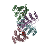
| Title | Crystal structure of Mycobacterium tuberculosis CYP126A1 in complex with 1-(3-(1H-imidazol-1-yl)propyl)-3-((3s,5s,7s)-adamantan-1-yl)urea | ||||||
 Components Components | Putative cytochrome P450 126 | ||||||
 Keywords Keywords |  OXIDOREDUCTASE / monoxygenase / cytochrome P450 OXIDOREDUCTASE / monoxygenase / cytochrome P450 | ||||||
| Function / homology |  Function and homology informationOxidoreductases; Acting on paired donors, with incorporation or reduction of molecular oxygen / oxidoreductase activity, acting on paired donors, with incorporation or reduction of molecular oxygen / monooxygenase activity / iron ion binding / heme binding Function and homology informationOxidoreductases; Acting on paired donors, with incorporation or reduction of molecular oxygen / oxidoreductase activity, acting on paired donors, with incorporation or reduction of molecular oxygen / monooxygenase activity / iron ion binding / heme bindingSimilarity search - Function | ||||||
| Biological species |   Mycobacterium tuberculosis (bacteria) Mycobacterium tuberculosis (bacteria) | ||||||
| Method | X-RAY DIFFRACTION / SYNCHROTRON / MOLECULAR REPLACEMENT / Resolution: 1.58 Å | ||||||
 Authors Authors | Levy, C. / Munro, A.W. / Leys, D. | ||||||
 Citation Citation | Journal: J. Biol. Chem. / Year: 2017 Title: Structural Characterization and Ligand/Inhibitor Identification Provide Functional Insights into the Mycobacterium tuberculosis Cytochrome P450 CYP126A1. Authors: Chenge, J.T. / Duyet, L.V. / Swami, S. / McLean, K.J. / Kavanagh, M.E. / Coyne, A.G. / Rigby, S.E. / Cheesman, M.R. / Girvan, H.M. / Levy, C.W. / Rupp, B. / von Kries, J.P. / Abell, C. / Leys, D. / Munro, A.W. | ||||||
| History |
|
- Structure visualization
| Structure viewer | Molecule: MolmilJmol/JSmol |
|---|
- Downloads & links
-Download
| PDBx/mmCIF format | 5li7.cif.gz | 192.6 KB | Display | PDBx/mmCIF format |
|---|---|---|---|---|
| PDB format | pdb5li7.ent.gz | 148.2 KB | Display | PDB format |
| PDBx/mmJSON format | 5li7.json.gz | Tree view | PDBx/mmJSON format | |
| Others |  Other downloads Other downloads |
-Validation report
| Arichive directory | https://data.pdbj.org/pub/pdb/validation_reports/li/5li7ftp://data.pdbj.org/pub/pdb/validation_reports/li/5li7 | HTTPS FTP |
|---|
-Related structure data
| Related structure data |  5li6C  5li8C  5lieC  1oxaS S: Starting model for refinement C: citing same article ( |
|---|---|
| Similar structure data |


-Links
 PDBj
PDBj
- Assembly
| Deposited unit | 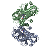
| ||||||||
|---|---|---|---|---|---|---|---|---|---|
| 1 |
| ||||||||
| Unit cell |
|
-Components
| #1: Protein | Mass: 46010.891 Da / Num. of mol.: 2 Source method: isolated from a genetically manipulated source Source: (gene. exp.) Mycobacterium tuberculosis (strain CDC 1551 / Oshkosh) (bacteria)Gene: cyp126, MT0802 / Production host: Escherichia coli (E. coli)References: UniProt: P9WPN8, Oxidoreductases; Acting on paired donors, with incorporation or reduction of molecular oxygen#2: Chemical | Heme B  Mass: 616.487 Da / Num. of mol.: 2 / Source method: obtained synthetically / Formula: C34H32FeN4O4 Mass: 616.487 Da / Num. of mol.: 2 / Source method: obtained synthetically / Formula: C34H32FeN4O4#3: Chemical |   Mass: 302.415 Da / Num. of mol.: 2 / Source method: obtained synthetically / Formula: C17H26N4O Mass: 302.415 Da / Num. of mol.: 2 / Source method: obtained synthetically / Formula: C17H26N4O#4: Water | ChemComp-HOH / | Water Mass: 18.015 Da / Num. of mol.: 998 / Source method: isolated from a natural source / Formula: H2O Mass: 18.015 Da / Num. of mol.: 998 / Source method: isolated from a natural source / Formula: H2O |
|---|
-Experimental details
-Experiment
| Experiment | Method: X-RAY DIFFRACTION / Number of used crystals: 1 |
|---|
- Sample preparation
| Crystal | Density Matthews: 2.54 Å3/Da / Density % sol: 51.67 % |
|---|---|
| Crystal grow | Temperature: 277 K / Method: vapor diffusion, sitting drop Details: 0.2M sodium sulphate, 0.1 M Hepes pH 7.5 and 20% PEG 3350 |
-Data collection
| Diffraction | Mean temperature: 100 K |
|---|---|
| Diffraction source | Source: SYNCHROTRON / Site: Diamond  / Beamline: I03 / Wavelength: 0.976 Å / Beamline: I03 / Wavelength: 0.976 Å |
| Detector | Type: DECTRIS PILATUS 300K / Detector: PIXEL / Date: Jan 19, 2011 |
| Radiation | Protocol: SINGLE WAVELENGTH / Monochromatic (M) / Laue (L): M / Scattering type: x-ray |
| Radiation wavelength | Wavelength: 0.976 Å / Relative weight: 1 |
| Reflection | Resolution: 1.58→57.31 Å / Num. obs: 122489 / % possible obs: 99.8 % / Redundancy: 7.3 % / Rmerge(I) obs: 0.024 / Net I/σ(I): 20.1 |
| Reflection shell | Resolution: 1.58→1.62 Å / Redundancy: 5.4 % / Rmerge(I) obs: 0.829 / Mean I/σ(I) obs: 2.6 / % possible all: 100 |
- Processing
| Software |
| ||||||||||||||||||||||||||||||||||||||||||||||||||||||||||||||||||||||||||||||||||||||||||||||||||||||||||||||||||||||||||||||||||||||||||||||||||||||||||||||||||||||||||||||||||||||
|---|---|---|---|---|---|---|---|---|---|---|---|---|---|---|---|---|---|---|---|---|---|---|---|---|---|---|---|---|---|---|---|---|---|---|---|---|---|---|---|---|---|---|---|---|---|---|---|---|---|---|---|---|---|---|---|---|---|---|---|---|---|---|---|---|---|---|---|---|---|---|---|---|---|---|---|---|---|---|---|---|---|---|---|---|---|---|---|---|---|---|---|---|---|---|---|---|---|---|---|---|---|---|---|---|---|---|---|---|---|---|---|---|---|---|---|---|---|---|---|---|---|---|---|---|---|---|---|---|---|---|---|---|---|---|---|---|---|---|---|---|---|---|---|---|---|---|---|---|---|---|---|---|---|---|---|---|---|---|---|---|---|---|---|---|---|---|---|---|---|---|---|---|---|---|---|---|---|---|---|---|---|---|---|
| Refinement | Method to determine structure: MOLECULAR REPLACEMENT Starting model: 1OXA Resolution: 1.58→57.31 Å / Cor.coef. Fo:Fc: 0.964 / Cor.coef. Fo:Fc free: 0.95 / SU B: 1.425 / SU ML: 0.051 / Cross valid method: THROUGHOUT / ESU R: 0.075 / ESU R Free: 0.079 / Details: HYDROGENS HAVE BEEN ADDED IN THE RIDING POSITIONS
| ||||||||||||||||||||||||||||||||||||||||||||||||||||||||||||||||||||||||||||||||||||||||||||||||||||||||||||||||||||||||||||||||||||||||||||||||||||||||||||||||||||||||||||||||||||||
| Solvent computation | Ion probe radii: 0.8 Å / Shrinkage radii: 0.8 Å / VDW probe radii: 1.4 Å | ||||||||||||||||||||||||||||||||||||||||||||||||||||||||||||||||||||||||||||||||||||||||||||||||||||||||||||||||||||||||||||||||||||||||||||||||||||||||||||||||||||||||||||||||||||||
| Displacement parameters | Biso mean: 16.426 Å2
| ||||||||||||||||||||||||||||||||||||||||||||||||||||||||||||||||||||||||||||||||||||||||||||||||||||||||||||||||||||||||||||||||||||||||||||||||||||||||||||||||||||||||||||||||||||||
| Refinement step | Cycle: 1 / Resolution: 1.58→57.31 Å
| ||||||||||||||||||||||||||||||||||||||||||||||||||||||||||||||||||||||||||||||||||||||||||||||||||||||||||||||||||||||||||||||||||||||||||||||||||||||||||||||||||||||||||||||||||||||
| Refine LS restraints |
|