 Movie
Movie Controller


+ Open data

- Basic information
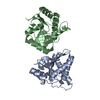
| Entry | Database: PDB / ID: 5lc4 | ||||||
|---|---|---|---|---|---|---|---|
| Title | Xray structure of mouse FAM3C ILEI dimer | ||||||
 Components Components | Protein FAM3C | ||||||
 Keywords Keywords |  SIGNALING PROTEIN SIGNALING PROTEIN | ||||||
| Function / homology | FAM3C (ILEI) domain / FAM3 family / ILEI/PANDER domain / Interleukin-like EMT inducer / Platelet degranulation / cytoplasmic vesicle / Golgi apparatus / extracellular space / Protein FAM3C Function and homology information Function and homology information | ||||||
| Biological species |  Mus musculus (house mouse) Mus musculus (house mouse) | ||||||
| Method | X-RAY DIFFRACTION / SYNCHROTRON / MOLECULAR REPLACEMENT / Resolution: 1.84 Å | ||||||
 Authors Authors | Johansson, P. / Jansson, A. | ||||||
 Citation Citation | Journal: J. Biol. Chem. / Year: 2017 Title: The interleukin-like epithelial-mesenchymal transition inducer ILEI exhibits a non-interleukin-like fold and is active as a domain-swapped dimer. Authors: Jansson, A.M. / Csiszar, A. / Maier, J. / Nystrom, A.C. / Ax, E. / Johansson, P. / Schiavone, L.H. | ||||||
| History |
|
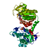
- Structure visualization
| Structure viewer | Molecule: MolmilJmol/JSmol |
|---|
- Downloads & links
-Download
| PDBx/mmCIF format | 5lc4.cif.gz | 278 KB | Display | PDBx/mmCIF format |
|---|---|---|---|---|
| PDB format | pdb5lc4.ent.gz | 224.5 KB | Display | PDB format |
| PDBx/mmJSON format | 5lc4.json.gz | Tree view | PDBx/mmJSON format | |
| Others |  Other downloads Other downloads |
-Validation report
| Arichive directory | https://data.pdbj.org/pub/pdb/validation_reports/lc/5lc4ftp://data.pdbj.org/pub/pdb/validation_reports/lc/5lc4 | HTTPS FTP |
|---|
-Related structure data
| Related structure data |  5lc2C  5lc3C 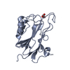 2yopS S: Starting model for refinement C: citing same article ( |
|---|---|
| Similar structure data |


-Links
 PDBj
PDBj
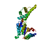
- Assembly
| Deposited unit | 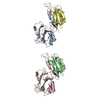
| ||||||||
|---|---|---|---|---|---|---|---|---|---|
| 1 | 
| ||||||||
| 2 | 
| ||||||||
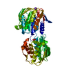
| Unit cell |
|
-Components
| #1: Protein | Mass: 22707.094 Da / Num. of mol.: 4 Source method: isolated from a genetically manipulated source Source: (gene. exp.) Mus musculus (house mouse) / Gene: Fam3c, D6Wsu176e, Ilei / Cell line (production host): Expi293 / Production host:  Homo sapiens (human) / References: UniProt: Q91VU0 Homo sapiens (human) / References: UniProt: Q91VU0#2: Water | ChemComp-HOH / | Water Mass: 18.015 Da / Num. of mol.: 423 / Source method: isolated from a natural source / Formula: H2O Mass: 18.015 Da / Num. of mol.: 423 / Source method: isolated from a natural source / Formula: H2O |
|---|
-Experimental details
-Experiment
| Experiment | Method: X-RAY DIFFRACTION / Number of used crystals: 1 |
|---|
- Sample preparation
| Crystal | Density Matthews: 2.67 Å3/Da / Density % sol: 53.88 % |
|---|---|
| Crystal grow | Temperature: 293 K / Method: vapor diffusion, sitting drop / pH: 6.5 Details: ILEI dimer 10 mg/mL + 3.3 M (NH4)2SO4, 1% MPD, 100 mM MES pH 6.5 |
-Data collection
| Diffraction | Mean temperature: 100 K |
|---|---|
| Diffraction source | Source: SYNCHROTRON / Site: ESRF  / Beamline: ID29 / Wavelength: 0.979 Å / Beamline: ID29 / Wavelength: 0.979 Å |
| Detector | Type: DECTRIS PILATUS 6M / Detector: PIXEL / Date: Jun 16, 2014 |
| Radiation | Protocol: SINGLE WAVELENGTH / Monochromatic (M) / Laue (L): M / Scattering type: x-ray |
| Radiation wavelength | Wavelength: 0.979 Å / Relative weight: 1 |
| Reflection | Resolution: 1.84→48.5 Å / Num. obs: 69589 / % possible obs: 98.4 % / Redundancy: 3.4 % / Biso Wilson estimate: 33.75 Å2 / Rmerge(I) obs: 0.078 / Net I/σ(I): 8.2 |
| Reflection shell | Resolution: 1.84→1.89 Å |
- Processing
| Software |
| |||||||||||||||||||||||||||||||||||||||||||||||||||||||||||||||||||||||||||||||||||||||||||||||||||||||||||||||||||||||||||||
|---|---|---|---|---|---|---|---|---|---|---|---|---|---|---|---|---|---|---|---|---|---|---|---|---|---|---|---|---|---|---|---|---|---|---|---|---|---|---|---|---|---|---|---|---|---|---|---|---|---|---|---|---|---|---|---|---|---|---|---|---|---|---|---|---|---|---|---|---|---|---|---|---|---|---|---|---|---|---|---|---|---|---|---|---|---|---|---|---|---|---|---|---|---|---|---|---|---|---|---|---|---|---|---|---|---|---|---|---|---|---|---|---|---|---|---|---|---|---|---|---|---|---|---|---|---|---|
| Refinement | Method to determine structure: MOLECULAR REPLACEMENT Starting model: 2YOP Resolution: 1.84→48.48 Å / Cor.coef. Fo:Fc: 0.935 / Cor.coef. Fo:Fc free: 0.926 / SU R Cruickshank DPI: 0.136 / Cross valid method: THROUGHOUT / σ(F): 0 / SU R Blow DPI: 0.137 / SU Rfree Blow DPI: 0.121 / SU Rfree Cruickshank DPI: 0.121
| |||||||||||||||||||||||||||||||||||||||||||||||||||||||||||||||||||||||||||||||||||||||||||||||||||||||||||||||||||||||||||||
| Displacement parameters | Biso mean: 39.6 Å2
| |||||||||||||||||||||||||||||||||||||||||||||||||||||||||||||||||||||||||||||||||||||||||||||||||||||||||||||||||||||||||||||
| Refine analyze | Luzzati coordinate error obs: 0.29 Å | |||||||||||||||||||||||||||||||||||||||||||||||||||||||||||||||||||||||||||||||||||||||||||||||||||||||||||||||||||||||||||||
| Refinement step | Cycle: LAST / Resolution: 1.84→48.48 Å
| |||||||||||||||||||||||||||||||||||||||||||||||||||||||||||||||||||||||||||||||||||||||||||||||||||||||||||||||||||||||||||||
| Refine LS restraints |
| |||||||||||||||||||||||||||||||||||||||||||||||||||||||||||||||||||||||||||||||||||||||||||||||||||||||||||||||||||||||||||||
| LS refinement shell | Resolution: 1.84→1.89 Å / Rfactor Rfree error: 0 / Total num. of bins used: 20
| |||||||||||||||||||||||||||||||||||||||||||||||||||||||||||||||||||||||||||||||||||||||||||||||||||||||||||||||||||||||||||||
| Refinement TLS params. | Method: refined / Refine-ID: X-RAY DIFFRACTION
| |||||||||||||||||||||||||||||||||||||||||||||||||||||||||||||||||||||||||||||||||||||||||||||||||||||||||||||||||||||||||||||
| Refinement TLS group |
|
