 Movie
Movie Controller

[English] 日本語
 Yorodumi
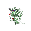
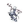
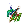
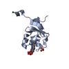
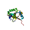
- PDB-5lam: Refined 3D NMR structure of the cytoplasmic rhodanese domain of t... -
+ Open data

- Basic information
| Entry | Database: PDB / ID: 5lam | ||||||||||||
|---|---|---|---|---|---|---|---|---|---|---|---|---|---|
| Title | Refined 3D NMR structure of the cytoplasmic rhodanese domain of the inner membrane protein YgaP from Escherichia coli | ||||||||||||
 Components Components | Inner membrane protein YgaP | ||||||||||||
 Keywords Keywords |  MEMBRANE PROTEIN / Rhodanese domain of YgaP / PROTEIN MEMBRANE PROTEIN / Rhodanese domain of YgaP / PROTEIN | ||||||||||||
| Function / homology |  Function and homology information Function and homology information | ||||||||||||
| Biological species |  Escherichia coli (E. coli) Escherichia coli (E. coli) | ||||||||||||
| Method | SOLUTION NMR / Energy refinement | ||||||||||||
 Authors Authors | Eichmann, C. / Tzitzilonis, C. / Nakamura, T. / Maslennikov, I. / Kwiatkowski, W. / Choe, S. / Lipton, S.A. / Guntert, P. / Riek, R. | ||||||||||||
| Funding support |  United States, 3items United States, 3items
| ||||||||||||
 Citation Citation | Journal: J.Mol.Biol. / Year: 2016 Title: S-Nitrosylation Induces Structural and Dynamical Changes in a Rhodanese Family Protein. Authors: Eichmann, C. / Tzitzilonis, C. / Nakamura, T. / Kwiatkowski, W. / Maslennikov, I. / Choe, S. / Lipton, S.A. / Riek, R. | ||||||||||||
| History |
|
- Structure visualization
| Structure viewer | Molecule: MolmilJmol/JSmol |
|---|
- Downloads & links
-Download
| PDBx/mmCIF format | 5lam.cif.gz | 641.6 KB | Display | PDBx/mmCIF format |
|---|---|---|---|---|
| PDB format | pdb5lam.ent.gz | 536.9 KB | Display | PDB format |
| PDBx/mmJSON format | 5lam.json.gz | Tree view | PDBx/mmJSON format | |
| Others |  Other downloads Other downloads |
-Validation report
| Arichive directory | https://data.pdbj.org/pub/pdb/validation_reports/la/5lamftp://data.pdbj.org/pub/pdb/validation_reports/la/5lam | HTTPS FTP |
|---|
-Related structure data
| Related structure data | 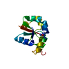 5hblC 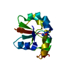 5hboC  5hbpC  5hbqC 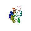 5laoC C: citing same article ( |
|---|---|
| Similar structure data | |
| Other databases |


-Links
 PDBj
PDBj- Assembly
| Deposited unit | 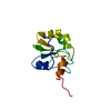
| |||||||||
|---|---|---|---|---|---|---|---|---|---|---|
| 1 |
| |||||||||
| NMR ensembles |
|
-Components
| #1: Protein | Mass: 11716.311 Da / Num. of mol.: 1 Source method: isolated from a genetically manipulated source Source: (gene. exp.) Escherichia coli (strain K12) (bacteria)Strain: K12 / Gene: ygaP, b2668, JW2643 / Production host: Escherichia coli BL21(DE3) (bacteria) / Variant (production host): pLysS Star / References: UniProt: P55734 |
|---|
-Experimental details
-Experiment
| Experiment | Method: SOLUTION NMR | ||||||||||||||||||||||||||||||||||||||||||
|---|---|---|---|---|---|---|---|---|---|---|---|---|---|---|---|---|---|---|---|---|---|---|---|---|---|---|---|---|---|---|---|---|---|---|---|---|---|---|---|---|---|---|---|
| NMR experiment |
|
- Sample preparation
| Details | Type: solution Contents: 1 mM [U-99% 13C; U-99% 15N] Rhodanese domain, 95% H2O/5% D2O Label: Rhodanese / Solvent system: 95% H2O/5% D2O |
|---|---|
| Sample | Conc.: 1 mM / Component: Rhodanese domain / Isotopic labeling: [U-99% 13C; U-99% 15N] |
| Sample conditions | Ionic strength: 0 mM / Label: Rhodanese / pH: 7 / Pressure: AMBIENT Pa / Temperature: 303.15 K |
-NMR measurement
| NMR spectrometer | Type: Bruker AVANCE III / Manufacturer: Bruker / Model: AVANCE III / Field strength: 700 MHz |
|---|
- Processing
| NMR software |
| ||||||||||||||||
|---|---|---|---|---|---|---|---|---|---|---|---|---|---|---|---|---|---|
| Refinement | Method: Energy refinement / Software ordinal: 1 | ||||||||||||||||
| NMR representative | Selection criteria: lowest energy | ||||||||||||||||
| NMR ensemble | Conformer selection criteria: structures with the least restraint violations Conformers calculated total number: 100 / Conformers submitted total number: 20 |
