 Movie
Movie Controller


+ Open data

- Basic information
| Entry | Database: PDB / ID: 5l8i | ||||||
|---|---|---|---|---|---|---|---|
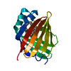
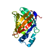
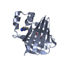
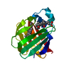
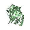
| Title | crystal structure of human FABP6 apo-protein | ||||||
 Components Components | Gastrotropin | ||||||
 Keywords Keywords |  SIGNALING PROTEIN / FABP6 / Fatty acid binding protein 6 / Ileal bile acid binding protein / I-BABP / Ileal / Gastrotropin / Fragments SIGNALING PROTEIN / FABP6 / Fatty acid binding protein 6 / Ileal bile acid binding protein / I-BABP / Ileal / Gastrotropin / Fragments | ||||||
| Function / homology |  Function and homology information Function and homology informationNR1H2 & NR1H3 regulate gene expression to control bile acid homeostasis / Triglyceride catabolism / fatty acid transport / Recycling of bile acids and salts / fatty acid binding / lipid metabolic process / negative regulation of cell population proliferation / lipid binding / membrane / nucleus ...NR1H2 & NR1H3 regulate gene expression to control bile acid homeostasis / Triglyceride catabolism / fatty acid transport / Recycling of bile acids and salts / fatty acid binding / lipid metabolic process / negative regulation of cell population proliferation / lipid binding / membrane / nucleus / cytosol / cytoplasmSimilarity search - Function | ||||||
| Biological species |  Homo sapiens (human) Homo sapiens (human) | ||||||
| Method | X-RAY DIFFRACTION / SYNCHROTRON / MOLECULAR REPLACEMENT / Resolution: 1.88 Å | ||||||
 Authors Authors | Hendrick, A. / Mueller, I. / Leonard, P.M. / Davenport, R. / Mitchell, P. | ||||||
 Citation Citation | Journal: J.Med.Chem. / Year: 2016 Title: Identification and Investigation of Novel Binding Fragments in the Fatty Acid Binding Protein 6 (FABP6). Authors: Hendrick, A.G. / Muller, I. / Willems, H. / Leonard, P.M. / Irving, S. / Davenport, R. / Ito, T. / Reeves, J. / Wright, S. / Allen, V. / Wilkinson, S. / Heffron, H. / Bazin, R. / Turney, J. / Mitchell, P.J. | ||||||
| History |
|
- Structure visualization
| Structure viewer | Molecule: MolmilJmol/JSmol |
|---|
- Downloads & links
-Download
| PDBx/mmCIF format | 5l8i.cif.gz | 157.6 KB | Display | PDBx/mmCIF format |
|---|---|---|---|---|
| PDB format | pdb5l8i.ent.gz | 124.6 KB | Display | PDB format |
| PDBx/mmJSON format | 5l8i.json.gz | Tree view | PDBx/mmJSON format | |
| Others |  Other downloads Other downloads |
-Validation report
| Arichive directory | https://data.pdbj.org/pub/pdb/validation_reports/l8/5l8iftp://data.pdbj.org/pub/pdb/validation_reports/l8/5l8i | HTTPS FTP |
|---|
-Related structure data
| Related structure data |  5l8nC  5l8oC  3exlS S: Starting model for refinement C: citing same article ( |
|---|---|
| Similar structure data |


-Links
 PDBj
PDBj


- Assembly
| Deposited unit | 
| ||||||||
|---|---|---|---|---|---|---|---|---|---|
| 1 | 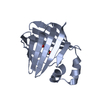
| ||||||||
| 2 | 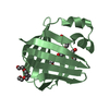
| ||||||||
| 3 | 
| ||||||||
| Unit cell |
|
-Components
-Protein , 1 types, 3 molecules ABC
| #1: Protein | Mass: 14389.235 Da / Num. of mol.: 3 Source method: isolated from a genetically manipulated source Source: (gene. exp.) Homo sapiens (human) / Gene: FABP6, ILBP, ILLBP / Production host:  Escherichia coli (E. coli) / References: UniProt: P51161 Escherichia coli (E. coli) / References: UniProt: P51161 |
|---|
-Non-polymers , 5 types, 63 molecules 


| #2: Chemical | ChemComp-PGE / Polyethylene glycol Mass: 150.173 Da / Num. of mol.: 1 / Source method: obtained synthetically / Formula: C6H14O4 Mass: 150.173 Da / Num. of mol.: 1 / Source method: obtained synthetically / Formula: C6H14O4 |
|---|---|
| #3: Chemical | ChemComp-P33 / Polyethylene glycol Mass: 326.383 Da / Num. of mol.: 1 / Source method: obtained synthetically / Formula: C14H30O8 / Comment: precipitant*YM Mass: 326.383 Da / Num. of mol.: 1 / Source method: obtained synthetically / Formula: C14H30O8 / Comment: precipitant*YM |
| #4: Chemical | ChemComp-PEG / Diethylene glycol Mass: 106.120 Da / Num. of mol.: 1 / Source method: obtained synthetically / Formula: C4H10O3 Mass: 106.120 Da / Num. of mol.: 1 / Source method: obtained synthetically / Formula: C4H10O3 |
| #5: Chemical | ChemComp-PG4 / Polyethylene glycol Mass: 194.226 Da / Num. of mol.: 1 / Source method: obtained synthetically / Formula: C8H18O5 / Comment: precipitant*YM Mass: 194.226 Da / Num. of mol.: 1 / Source method: obtained synthetically / Formula: C8H18O5 / Comment: precipitant*YM |
| #6: Water | ChemComp-HOH / WaterMass: 18.015 Da / Num. of mol.: 59 / Source method: isolated from a natural source / Formula: H2O |
-Experimental details
-Experiment
| Experiment | Method: X-RAY DIFFRACTION / Number of used crystals: 1 |
|---|
- Sample preparation
| Crystal | Density Matthews: 2.04 Å3/Da / Density % sol: 39.78 % |
|---|---|
| Crystal grow | Temperature: 293.5 K / Method: vapor diffusion, hanging drop / pH: 5.5 / Details: 41% w/v PEG 3350 and 0.1 M Bis-tris pH 5.5 |
-Data collection
| Diffraction | Mean temperature: 100 K |
|---|---|
| Diffraction source | Source: SYNCHROTRON / Site: Diamond  / Beamline: I02 / Wavelength: 0.97623 Å / Beamline: I02 / Wavelength: 0.97623 Å |
| Detector | Type: DECTRIS PILATUS 6M / Detector: PIXEL / Date: Aug 8, 2014 |
| Radiation | Protocol: SINGLE WAVELENGTH / Monochromatic (M) / Laue (L): M / Scattering type: x-ray |
| Radiation wavelength | Wavelength: 0.97623 Å / Relative weight: 1 |
| Reflection | Resolution: 1.88→58.58 Å / Num. obs: 28093 / % possible obs: 99 % / Redundancy: 3.2 % / Biso Wilson estimate: 34.5 Å2 / CC1/2: 0.994 / Rmerge(I) obs: 0.081 / Net I/σ(I): 5.5 |
| Reflection shell | Resolution: 1.88→1.96 Å / Redundancy: 2.8 % / Rmerge(I) obs: 0.627 / Mean I/σ(I) obs: 1.5 / % possible all: 95.6 |
- Processing
| Software |
| ||||||||||||||||||||||||||||||||||||||||||||||||||||||||||||||||||||||||||||||||||||||||||||||||||||||||||||||||||||||||||||||||||||||||||||||||||||||||||||||||||||||||||||||||||||||
|---|---|---|---|---|---|---|---|---|---|---|---|---|---|---|---|---|---|---|---|---|---|---|---|---|---|---|---|---|---|---|---|---|---|---|---|---|---|---|---|---|---|---|---|---|---|---|---|---|---|---|---|---|---|---|---|---|---|---|---|---|---|---|---|---|---|---|---|---|---|---|---|---|---|---|---|---|---|---|---|---|---|---|---|---|---|---|---|---|---|---|---|---|---|---|---|---|---|---|---|---|---|---|---|---|---|---|---|---|---|---|---|---|---|---|---|---|---|---|---|---|---|---|---|---|---|---|---|---|---|---|---|---|---|---|---|---|---|---|---|---|---|---|---|---|---|---|---|---|---|---|---|---|---|---|---|---|---|---|---|---|---|---|---|---|---|---|---|---|---|---|---|---|---|---|---|---|---|---|---|---|---|---|---|
| Refinement | Method to determine structure: MOLECULAR REPLACEMENT Starting model: 3exl Resolution: 1.88→58.58 Å / Cor.coef. Fo:Fc: 0.937 / Cor.coef. Fo:Fc free: 0.919 / SU B: 12.462 / SU ML: 0.169 / Cross valid method: THROUGHOUT / ESU R: 0.203 / ESU R Free: 0.175 / Details: HYDROGENS HAVE BEEN ADDED IN THE RIDING POSITIONS
| ||||||||||||||||||||||||||||||||||||||||||||||||||||||||||||||||||||||||||||||||||||||||||||||||||||||||||||||||||||||||||||||||||||||||||||||||||||||||||||||||||||||||||||||||||||||
| Solvent computation | Ion probe radii: 0.8 Å / Shrinkage radii: 0.8 Å / VDW probe radii: 1.2 Å | ||||||||||||||||||||||||||||||||||||||||||||||||||||||||||||||||||||||||||||||||||||||||||||||||||||||||||||||||||||||||||||||||||||||||||||||||||||||||||||||||||||||||||||||||||||||
| Displacement parameters | Biso mean: 38.573 Å2
| ||||||||||||||||||||||||||||||||||||||||||||||||||||||||||||||||||||||||||||||||||||||||||||||||||||||||||||||||||||||||||||||||||||||||||||||||||||||||||||||||||||||||||||||||||||||
| Refinement step | Cycle: 1 / Resolution: 1.88→58.58 Å
| ||||||||||||||||||||||||||||||||||||||||||||||||||||||||||||||||||||||||||||||||||||||||||||||||||||||||||||||||||||||||||||||||||||||||||||||||||||||||||||||||||||||||||||||||||||||
| Refine LS restraints |
|