 Movie
Movie Controller

[English] 日本語
 Yorodumi
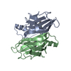
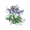
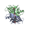
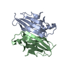
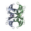
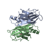
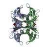
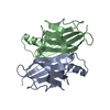
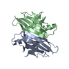
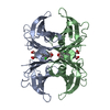
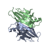
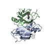
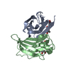
- PDB-5l4m: Crystal Structure of Human Transthyretin in Complex with 3,5,6-Tr... -
+ Open data

- Basic information
| Entry | Database: PDB / ID: 5l4m | ||||||
|---|---|---|---|---|---|---|---|
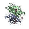
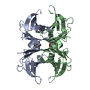
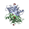
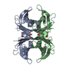
| Title | Crystal Structure of Human Transthyretin in Complex with 3,5,6-Trichloro-2-pyridinyloxyacetic acid (Triclopyr) | ||||||
 Components Components | Transthyretin | ||||||
 Keywords Keywords | TRANSPORT PROTEIN / thyroxine binding / 3 / 5 / 6-Trichloro-2-pyridinyloxyacetic acid (Triclopyr) complex | ||||||
| Function / homology |  Function and homology information Function and homology informationRetinoid cycle disease events / The canonical retinoid cycle in rods (twilight vision) / thyroid hormone binding / purine nucleobase metabolic process / Non-integrin membrane-ECM interactions / Retinoid metabolism and transport / hormone activity / azurophil granule lumen / Amyloid fiber formation / Neutrophil degranulation ...Retinoid cycle disease events / The canonical retinoid cycle in rods (twilight vision) / thyroid hormone binding / purine nucleobase metabolic process / Non-integrin membrane-ECM interactions / Retinoid metabolism and transport / hormone activity / azurophil granule lumen / Amyloid fiber formation / Neutrophil degranulation / extracellular space / extracellular exosome / extracellular region / identical protein bindingSimilarity search - Function | ||||||
| Biological species |  Homo sapiens (human) Homo sapiens (human) | ||||||
| Method | X-RAY DIFFRACTION / MOLECULAR REPLACEMENT / Resolution: 1.581 Å | ||||||
 Authors Authors | Grundstrom, C. / Hall, M. / Zhang, J. / Olofsson, A. / Andersson, P. / Sauer-Eriksson, A.E. | ||||||
| Funding support |  Sweden, 1items Sweden, 1items
| ||||||
 Citation Citation | Journal: Environ. Sci. Technol. / Year: 2016 Title: Structure-Based Virtual Screening Protocol for in Silico Identification of Potential Thyroid Disrupting Chemicals Targeting Transthyretin. Authors: Zhang, J. / Begum, A. / Brannstrom, K. / Grundstrom, C. / Iakovleva, I. / Olofsson, A. / Sauer-Eriksson, A.E. / Andersson, P.L. | ||||||
| History |
|
- Structure visualization
| Structure viewer | Molecule: MolmilJmol/JSmol |
|---|
- Downloads & links
-Download
| PDBx/mmCIF format | 5l4m.cif.gz | 152.7 KB | Display | PDBx/mmCIF format |
|---|---|---|---|---|
| PDB format | pdb5l4m.ent.gz | 123.2 KB | Display | PDB format |
| PDBx/mmJSON format | 5l4m.json.gz | Tree view | PDBx/mmJSON format | |
| Others |  Other downloads Other downloads |
-Validation report
| Arichive directory | https://data.pdbj.org/pub/pdb/validation_reports/l4/5l4mftp://data.pdbj.org/pub/pdb/validation_reports/l4/5l4m | HTTPS FTP |
|---|
-Related structure data
| Related structure data | 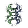 5jidC  5jimC 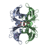 5jiqC  5l4fC 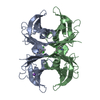 5l4iC  5l4jC 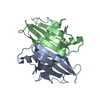 1f41S S: Starting model for refinement C: citing same article ( |
|---|---|
| Similar structure data |


-Links
 PDBj
PDBj


- Assembly
| Deposited unit | 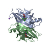
| ||||||||||||||||||
|---|---|---|---|---|---|---|---|---|---|---|---|---|---|---|---|---|---|---|---|
| 1 | 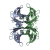
| ||||||||||||||||||
| Unit cell |
| ||||||||||||||||||
| Components on special symmetry positions |
|
-Components
| #1: Protein | / ATTR / Prealbumin / TBPA Mass: 13777.360 Da / Num. of mol.: 2 Source method: isolated from a genetically manipulated source Source: (gene. exp.) Homo sapiens (human) / Gene: TTR, PALB / Production host:  Escherichia coli (E. coli) / References: UniProt: P02766 Escherichia coli (E. coli) / References: UniProt: P02766#2: Chemical | Triclopyr  Mass: 256.471 Da / Num. of mol.: 2 / Source method: obtained synthetically / Formula: C7H4Cl3NO3 Mass: 256.471 Da / Num. of mol.: 2 / Source method: obtained synthetically / Formula: C7H4Cl3NO3#3: Chemical | ChemComp-NA / |   Mass: 22.990 Da / Num. of mol.: 1 / Source method: obtained synthetically / Formula: Na Mass: 22.990 Da / Num. of mol.: 1 / Source method: obtained synthetically / Formula: Na#4: Water | ChemComp-HOH / | Water Mass: 18.015 Da / Num. of mol.: 204 / Source method: isolated from a natural source / Formula: H2O Mass: 18.015 Da / Num. of mol.: 204 / Source method: isolated from a natural source / Formula: H2O |
|---|
-Experimental details
-Experiment
| Experiment | Method: X-RAY DIFFRACTION / Number of used crystals: 1 |
|---|
- Sample preparation
| Crystal | Density Matthews: 2.11 Å3/Da / Density % sol: 41.79 % |
|---|---|
| Crystal grow | Temperature: 291 K / Method: vapor diffusion, hanging drop / pH: 7.6 Details: Purified TTRwt was dialyzed against 10 mM sodium phosphate buffer with 100 mM KCl pH 7.6 and concentrated to 5 mg per ml. Triclopyr was added at 5 x molar excess to the protein. The ...Details: Purified TTRwt was dialyzed against 10 mM sodium phosphate buffer with 100 mM KCl pH 7.6 and concentrated to 5 mg per ml. Triclopyr was added at 5 x molar excess to the protein. The reservoir contained 1.3 to 1.6 M sodium citrate and 3.5 percent glycerol at pH 5.5. Drop size 3 plus 3 microliter |
-Data collection
| Diffraction | Mean temperature: 100 K |
|---|---|
| Diffraction source | Source: ROTATING ANODE / Type: BRUKER AXS MICROSTAR-H / Wavelength: 1.5418 Å |
| Detector | Type: Bruker Platinum 135 / Detector: CCD / Date: May 12, 2016 |
| Radiation | Protocol: SINGLE WAVELENGTH / Monochromatic (M) / Laue (L): M / Scattering type: x-ray |
| Radiation wavelength | Wavelength: 1.5418 Å / Relative weight: 1 |
| Reflection | Resolution: 1.58→27.3 Å / Num. obs: 31035 / % possible obs: 94.8 % / Redundancy: 3.1 % / Rmerge(I) obs: 0.081 / Net I/σ(I): 9.4 |
| Reflection shell | Resolution: 1.58→1.68 Å / Redundancy: 2.8 % / Rmerge(I) obs: 0.691 / Mean I/σ(I) obs: 1.4 / % possible all: 87.4 |
- Processing
| Software |
| |||||||||||||||||||||||||||||||||||||||||||||||||||||||||||||||||||||||||||||||||||||||||||||||||||||||||||||||||||||||||||||||||||||||||||||||||||
|---|---|---|---|---|---|---|---|---|---|---|---|---|---|---|---|---|---|---|---|---|---|---|---|---|---|---|---|---|---|---|---|---|---|---|---|---|---|---|---|---|---|---|---|---|---|---|---|---|---|---|---|---|---|---|---|---|---|---|---|---|---|---|---|---|---|---|---|---|---|---|---|---|---|---|---|---|---|---|---|---|---|---|---|---|---|---|---|---|---|---|---|---|---|---|---|---|---|---|---|---|---|---|---|---|---|---|---|---|---|---|---|---|---|---|---|---|---|---|---|---|---|---|---|---|---|---|---|---|---|---|---|---|---|---|---|---|---|---|---|---|---|---|---|---|---|---|---|---|
| Refinement | Method to determine structure: MOLECULAR REPLACEMENT Starting model: 1F41 Resolution: 1.581→27.282 Å / SU ML: 0.22 / Cross valid method: FREE R-VALUE / σ(F): 1.33 / Phase error: 22.09
| |||||||||||||||||||||||||||||||||||||||||||||||||||||||||||||||||||||||||||||||||||||||||||||||||||||||||||||||||||||||||||||||||||||||||||||||||||
| Solvent computation | Shrinkage radii: 0.9 Å / VDW probe radii: 1.11 Å | |||||||||||||||||||||||||||||||||||||||||||||||||||||||||||||||||||||||||||||||||||||||||||||||||||||||||||||||||||||||||||||||||||||||||||||||||||
| Refinement step | Cycle: LAST / Resolution: 1.581→27.282 Å
| |||||||||||||||||||||||||||||||||||||||||||||||||||||||||||||||||||||||||||||||||||||||||||||||||||||||||||||||||||||||||||||||||||||||||||||||||||
| Refine LS restraints |
| |||||||||||||||||||||||||||||||||||||||||||||||||||||||||||||||||||||||||||||||||||||||||||||||||||||||||||||||||||||||||||||||||||||||||||||||||||
| LS refinement shell |
|
