 Movie
Movie Controller

[English] 日本語
 Yorodumi
- PDB-5l0o: IQGAP1 calponin homology domain fragment (CHDF) mutant K161C unde... -
+ Open data

- Basic information
| Entry | Database: PDB / ID: 5l0o | ||||||
|---|---|---|---|---|---|---|---|
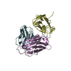
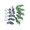
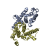
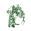
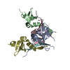
| Title | IQGAP1 calponin homology domain fragment (CHDF) mutant K161C under oxidizing conditions | ||||||
 Components Components | Ras GTPase-activating-like protein IQGAP1 | ||||||
 Keywords Keywords |  SIGNALING PROTEIN / Calponin homology / disulfide-bonded dimer SIGNALING PROTEIN / Calponin homology / disulfide-bonded dimer | ||||||
| Function / homology |  Function and homology information Function and homology informationnegative regulation of dephosphorylation / mitotic actomyosin contractile ring assembly actin filament organization / podocyte development / slit diaphragm / GTPase inhibitor activity / fibroblast migration / MAP-kinase scaffold activity / S100 protein binding / Nephrin family interactions / neuron projection extension ...negative regulation of dephosphorylation / mitotic actomyosin contractile ring assembly actin filament organization / podocyte development / slit diaphragm / GTPase inhibitor activity / fibroblast migration / MAP-kinase scaffold activity / S100 protein binding / Nephrin family interactions / neuron projection extension / RHOV GTPase cycle / cortical actin cytoskeleton / cellular response to platelet-derived growth factor stimulus / RHOC GTPase cycle / RHOQ GTPase cycle / phosphatidylinositol-3,4,5-trisphosphate binding / CDC42 GTPase cycle / RHOU GTPase cycle / platelet-derived growth factor receptor signaling pathway / lateral plasma membrane / RHOA GTPase cycle / RAC2 GTPase cycle / RHO GTPases activate IQGAPs / positive regulation of protein kinase activity / fibroblast growth factor receptor signaling pathway / regulation of cytokine production / cellular response to epidermal growth factor stimulus / ruffle / RAC1 GTPase cycle / regulation of mitotic cell cycle / cellular response to calcium ion / protein serine/threonine kinase activator activity / GTPase activator activity / secretory granule membrane / actin filament / Signaling by high-kinase activity BRAF mutants / MAP2K and MAPK activation / epidermal growth factor receptor signaling pathway / cytoplasmic side of plasma membrane / small GTPase binding / Glucagon-like Peptide-1 (GLP1) regulates insulin secretion / cytoplasmic ribonucleoprotein granule / Signaling by RAF1 mutants / Signaling by moderate kinase activity BRAF mutants / Paradoxical activation of RAF signaling by kinase inactive BRAF / Signaling downstream of RAS mutants / Signaling by BRAF and RAF1 fusions / actin filament binding / cell migration / cell cortex / midbody / growth cone / basolateral plasma membrane / protein phosphatase binding / microtubule / positive regulation of MAPK cascade / molecular adaptor activity / calmodulin binding / neuron projection / cadherin binding / ribonucleoprotein complex / apical plasma membrane / protein domain specific binding / axon / focal adhesion / calcium ion binding / Neutrophil degranulation / protein kinase binding / signal transduction / extracellular exosome / nucleus / plasma membrane / cytosol / cytoplasmSimilarity search - Function | ||||||
| Biological species |  Homo sapiens (human) Homo sapiens (human) | ||||||
| Method | X-RAY DIFFRACTION / MOLECULAR REPLACEMENT / Resolution: 2.36 Å | ||||||
 Authors Authors | Liu, J. / Worthylake, D. | ||||||
| Funding support |  United States, 1items United States, 1items
| ||||||
 Citation Citation | Journal: Biochemistry / Year: 2016 Title: The IQGAP1 N-Terminus Forms Dimers, and the Dimer Interface Is Required for Binding F-Actin and Calcium-Bound Calmodulin. Authors: Liu, J. / Kurella, V.B. / LeCour, L. / Vanagunas, T. / Worthylake, D.K. | ||||||
| History |
|
- Structure visualization
| Structure viewer | Molecule: MolmilJmol/JSmol |
|---|
- Downloads & links
-Download
| PDBx/mmCIF format | 5l0o.cif.gz | 142 KB | Display | PDBx/mmCIF format |
|---|---|---|---|---|
| PDB format | pdb5l0o.ent.gz | 112.7 KB | Display | PDB format |
| PDBx/mmJSON format | 5l0o.json.gz | Tree view | PDBx/mmJSON format | |
| Others |  Other downloads Other downloads |
-Validation report
| Arichive directory | https://data.pdbj.org/pub/pdb/validation_reports/l0/5l0oftp://data.pdbj.org/pub/pdb/validation_reports/l0/5l0o | HTTPS FTP |
|---|
-Related structure data
| Related structure data |  3i6xSC S: Starting model for refinement C: citing same article ( |
|---|---|
| Similar structure data |


-Links
 PDBj
PDBj


- Assembly
| Deposited unit | 
| ||||||||
|---|---|---|---|---|---|---|---|---|---|
| 1 | 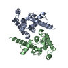
| ||||||||
| 2 | 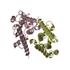
| ||||||||
| Unit cell |
| ||||||||
| Details | Dimer according to gel filtration, mutagenesis, shape complementary analysis |
-Components
| #1: Protein | Mass: 22361.402 Da / Num. of mol.: 4 / Mutation: K161C Source method: isolated from a genetically manipulated source Source: (gene. exp.) Homo sapiens (human) / Gene: IQGAP1, KIAA0051 / Production host:  Escherichia coli (E. coli) Escherichia coli (E. coli)Strain (production host): in Rosetta Gami 2(DE3) pLysS cells (EMD4Biosciences) References: UniProt: P46940 |
|---|
-Experimental details
-Experiment
| Experiment | Method: X-RAY DIFFRACTION / Number of used crystals: 1 |
|---|
- Sample preparation
| Crystal | Density Matthews: 2.96 Å3/Da / Density % sol: 58.44 % |
|---|---|
| Crystal grow | Temperature: 277 K / Method: vapor diffusion / pH: 8 Details: 18% PEG 3350, 10% ethylene glycol, 10% glucose, 100mM -160mM potassium acetate |
-Data collection
| Diffraction | Mean temperature: 100 K |
|---|---|
| Diffraction source | Source: ROTATING ANODE / Type: BRUKER AXS MICROSTAR / Wavelength: 1.5418 Å |
| Detector | Type: Bruker Platinum 135 / Detector: CCD / Date: Jun 25, 2014 |
| Radiation | Protocol: SINGLE WAVELENGTH / Monochromatic (M) / Laue (L): M / Scattering type: x-ray |
| Radiation wavelength | Wavelength: 1.5418 Å / Relative weight: 1 |
| Reflection | Resolution: 2.36→68.06 Å / Num. obs: 41528 / % possible obs: 98.4 % / Redundancy: 3.13 % / Rmerge(I) obs: 0.0847 / Net I/σ(I): 25.57 |
| Reflection shell | Resolution: 2.36→2.46 Å / Redundancy: 1.79 % / Rmerge(I) obs: 0.3504 / Mean I/σ(I) obs: 2.14 / % possible all: 94.8 |
- Processing
| Software |
| ||||||||||||||||||||
|---|---|---|---|---|---|---|---|---|---|---|---|---|---|---|---|---|---|---|---|---|---|
| Refinement | Method to determine structure: MOLECULAR REPLACEMENT Starting model: 3I6X Resolution: 2.36→15 Å / Data cutoff low absF: 0 / Cross valid method: FREE R-VALUE
| ||||||||||||||||||||
| Refinement step | Cycle: LAST / Resolution: 2.36→15 Å
|
