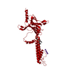
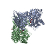
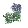
METAL TRANSPORT / METAL TRANSPORT Kir 2.2 K62W mutant structure
Function / homology
Function and homology information
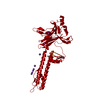
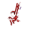
Activation of G protein gated Potassium channels / Classical Kir channels / Phase 4 - resting membrane potential / Inhibition of voltage gated Ca2+ channels via Gbeta/gamma subunits / inward rectifier potassium channel activity / regulation of monoatomic ion transmembrane transport / monoatomic ion channel complex / potassium ion import across plasma membrane / potassium ion transport / protein homotetramerization ...Activation of G protein gated Potassium channels / Classical Kir channels / Phase 4 - resting membrane potential / Inhibition of voltage gated Ca2+ channels via Gbeta/gamma subunits / inward rectifier potassium channel activity / regulation of monoatomic ion transmembrane transport / monoatomic ion channel complex / potassium ion import across plasma membrane / potassium ion transport / protein homotetramerization / membrane / identical protein binding / plasma membrane Similarity search - Function
Resolution: 2→91.39 Å / Cor.coef. Fo:Fc: 0.951 / Cor.coef. Fo:Fc free: 0.934 / SU B: 4.116 / SU ML: 0.109 / Cross valid method: THROUGHOUT / ESU R: 0.134 / ESU R Free: 0.127 / Stereochemistry target values: MAXIMUM LIKELIHOOD / Details: HYDROGENS HAVE BEEN ADDED IN THE RIDING POSITIONS
Rfactor
Num. reflection
% reflection
Selection details
Rfree
0.22667
1965
4.8 %
RANDOM
Rwork
0.19906
-
-
-
obs
0.20038
38705
99.13 %
-
Solvent computation
Ion probe radii: 0.8 Å / Shrinkage radii: 0.8 Å / VDW probe radii: 1.2 Å / Solvent model: MASK
 Movie
Movie Controller


 Open data
 Basic information
 Components
 Keywords
 Function and homology information
 inward rectifier potassium channel activity / regulation of monoatomic ion transmembrane transport / monoatomic ion channel complex / potassium ion import across plasma membrane / potassium ion transport / protein homotetramerization ...Activation of G protein gated Potassium channels / Classical Kir channels / Phase 4 - resting membrane potential / Inhibition of voltage gated Ca2+ channels via Gbeta/gamma subunits /
 Authors
 United States, 2items
  Citation
 Structure visualization
 Downloads & links
 Other downloads
 PDBj
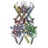
PDBj Assembly


 Mass: 39.098 Da / Num. of mol.: 9 / Source method: isolated from a natural source / Formula: K
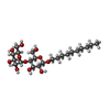
 Type: D-saccharide / Mass: 482.562 Da / Num. of mol.: 1
 Mass: 18.015 Da / Num. of mol.: 186 / Source method: isolated from a natural source / Formula: H2O
 Sample preparation
 Processing