 Movie
Movie Controller


+ Open data

- Basic information
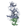
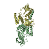
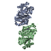
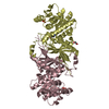
| Entry | Database: PDB / ID: 5kpt | ||||||
|---|---|---|---|---|---|---|---|
| Title | PANK3-AMPPNP complex | ||||||
 Components Components | Pantothenate kinase 3 | ||||||
 Keywords Keywords | TRANSFERASE / PANK / Substrate / Complex / Pantothenate kinase | ||||||
| Function / homology |  Function and homology information Function and homology informationCoenzyme A biosynthesis / vitamin binding / acetyl-CoA binding / pantothenate kinase / pantothenate kinase activity / coenzyme A biosynthetic process / phosphorylation / protein homodimerization activity / ATP binding / nucleus / cytosolSimilarity search - Function | ||||||
| Biological species |  Homo sapiens (human) Homo sapiens (human) | ||||||
| Method | X-RAY DIFFRACTION / SYNCHROTRON / MOLECULAR REPLACEMENT / Resolution: 2.301 Å | ||||||
 Authors Authors | White, S.W. / Yun, M. | ||||||
 Citation Citation | Journal: J.Biol.Chem. / Year: 2016 Title: Allosteric Regulation of Mammalian Pantothenate Kinase. Authors: Subramanian, C. / Yun, M.K. / Yao, J. / Sharma, L.K. / Lee, R.E. / White, S.W. / Jackowski, S. / Rock, C.O. | ||||||
| History |
|
- Structure visualization
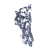
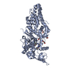
| Structure viewer | Molecule: MolmilJmol/JSmol |
|---|
- Downloads & links
-Download
| PDBx/mmCIF format | 5kpt.cif.gz | 154.5 KB | Display | PDBx/mmCIF format |
|---|---|---|---|---|
| PDB format | pdb5kpt.ent.gz | 117.9 KB | Display | PDB format |
| PDBx/mmJSON format | 5kpt.json.gz | Tree view | PDBx/mmJSON format | |
| Others |  Other downloads Other downloads |
-Validation report
| Arichive directory | https://data.pdbj.org/pub/pdb/validation_reports/kp/5kptftp://data.pdbj.org/pub/pdb/validation_reports/kp/5kpt | HTTPS FTP |
|---|
-Related structure data
| Related structure data |  5kprSC  5kpzC  5kq8C 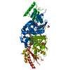 5kqdC S: Starting model for refinement C: citing same article ( |
|---|---|
| Similar structure data |


-Links
 PDBj
PDBj


- Assembly
| Deposited unit | 
| ||||||||
|---|---|---|---|---|---|---|---|---|---|
| 1 | 
| ||||||||
| Unit cell |
|
-Components
| #1: Protein | / hPanK3 / Pantothenic acid kinase 3 Mass: 42125.750 Da / Num. of mol.: 1 / Fragment: UNP resdiues 12-370 Source method: isolated from a genetically manipulated source Source: (gene. exp.) Homo sapiens (human) / Gene: PANK3 / Plasmid: PET28A / Production host:  Escherichia coli (E. coli) / Strain (production host): BL21(DE3) / References: UniProt: Q9H999, pantothenate kinase Escherichia coli (E. coli) / Strain (production host): BL21(DE3) / References: UniProt: Q9H999, pantothenate kinase |
|---|---|
| #2: Chemical | ChemComp-MG /   Mass: 24.305 Da / Num. of mol.: 1 / Source method: obtained synthetically / Formula: Mg Mass: 24.305 Da / Num. of mol.: 1 / Source method: obtained synthetically / Formula: Mg |
| #3: Chemical | ChemComp-ANP /   Mass: 506.196 Da / Num. of mol.: 1 / Source method: obtained synthetically / Formula: C10H17N6O12P3 / Comment: AMP-PNP, energy-carrying molecule analogue*YM Mass: 506.196 Da / Num. of mol.: 1 / Source method: obtained synthetically / Formula: C10H17N6O12P3 / Comment: AMP-PNP, energy-carrying molecule analogue*YM |
| #4: Chemical | ChemComp-EDO / Ethylene glycol  Mass: 62.068 Da / Num. of mol.: 1 / Source method: obtained synthetically / Formula: C2H6O2 Mass: 62.068 Da / Num. of mol.: 1 / Source method: obtained synthetically / Formula: C2H6O2 |
| #5: Water | ChemComp-HOH / Water Mass: 18.015 Da / Num. of mol.: 68 / Source method: isolated from a natural source / Formula: H2O Mass: 18.015 Da / Num. of mol.: 68 / Source method: isolated from a natural source / Formula: H2O |
-Experimental details
-Experiment
| Experiment | Method: X-RAY DIFFRACTION / Number of used crystals: 1 |
|---|
- Sample preparation
| Crystal | Density Matthews: 2.31 Å3/Da / Density % sol: 46.8 % |
|---|---|
| Crystal grow | Temperature: 291.15 K / Method: vapor diffusion, sitting drop / pH: 5.6 / Details: PEG 4000, ammonium acetate, citrate |
-Data collection
| Diffraction | Mean temperature: 100 K |
|---|---|
| Diffraction source | Source: SYNCHROTRON / Site: APS  / Beamline: 22-ID / Wavelength: 1 Å / Beamline: 22-ID / Wavelength: 1 Å |
| Detector | Type: RAYONIX MX300-HS / Detector: CCD / Date: Dec 16, 2015 |
| Radiation | Protocol: SINGLE WAVELENGTH / Monochromatic (M) / Laue (L): M / Scattering type: x-ray |
| Radiation wavelength | Wavelength: 1 Å / Relative weight: 1 |
| Reflection | Resolution: 2.3→39.642 Å / Num. obs: 16737 / % possible obs: 98.8 % / Observed criterion σ(I): -3 / Redundancy: 10.4 % / Rmerge(I) obs: 0.097 / Net I/σ(I): 22.4 |
| Reflection shell | Resolution: 2.3→2.36 Å / Redundancy: 5.8 % / Rmerge(I) obs: 0.578 / Mean I/σ(I) obs: 1.9 / CC1/2: 0.8 / % possible all: 89.9 |
- Processing
| Software |
| |||||||||||||||||||||||||||||||||||||||||||||||||||||||||||||||||||||||||||
|---|---|---|---|---|---|---|---|---|---|---|---|---|---|---|---|---|---|---|---|---|---|---|---|---|---|---|---|---|---|---|---|---|---|---|---|---|---|---|---|---|---|---|---|---|---|---|---|---|---|---|---|---|---|---|---|---|---|---|---|---|---|---|---|---|---|---|---|---|---|---|---|---|---|---|---|---|
| Refinement | Method to determine structure: MOLECULAR REPLACEMENT Starting model: 5KPR Resolution: 2.301→39.642 Å / SU ML: 0.27 / Cross valid method: THROUGHOUT / σ(F): 1.35 / Phase error: 24.05
| |||||||||||||||||||||||||||||||||||||||||||||||||||||||||||||||||||||||||||
| Solvent computation | Shrinkage radii: 0.9 Å / VDW probe radii: 1.11 Å | |||||||||||||||||||||||||||||||||||||||||||||||||||||||||||||||||||||||||||
| Displacement parameters | Biso mean: 42.42 Å2 | |||||||||||||||||||||||||||||||||||||||||||||||||||||||||||||||||||||||||||
| Refinement step | Cycle: LAST / Resolution: 2.301→39.642 Å
| |||||||||||||||||||||||||||||||||||||||||||||||||||||||||||||||||||||||||||
| Refine LS restraints |
| |||||||||||||||||||||||||||||||||||||||||||||||||||||||||||||||||||||||||||
| LS refinement shell |
| |||||||||||||||||||||||||||||||||||||||||||||||||||||||||||||||||||||||||||
| Refinement TLS params. | Method: refined / Refine-ID: X-RAY DIFFRACTION
| |||||||||||||||||||||||||||||||||||||||||||||||||||||||||||||||||||||||||||
| Refinement TLS group |
|
