 Movie
Movie Controller

[English] 日本語
 Yorodumi
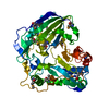
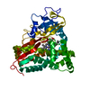
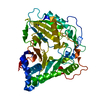
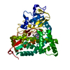
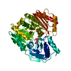
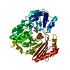
- PDB-5kjw: Crystal structure of Coleus blumei HCT in complex with 3-hydroxya... -
+ Open data

- Basic information
| Entry | Database: PDB / ID: 5kjw | ||||||||||||
|---|---|---|---|---|---|---|---|---|---|---|---|---|---|
| Title | Crystal structure of Coleus blumei HCT in complex with 3-hydroxyacetophenone | ||||||||||||
 Components Components | Hydroxycinnamoyl transferase | ||||||||||||
 Keywords Keywords |  TRANSFERASE / phenylpropanoid metabolism / BAHD / HCT / acyltransferase TRANSFERASE / phenylpropanoid metabolism / BAHD / HCT / acyltransferase | ||||||||||||
| Function / homology |  Function and homology informationshikimate O-hydroxycinnamoyltransferase / shikimate O-hydroxycinnamoyltransferase activity Function and homology informationshikimate O-hydroxycinnamoyltransferase / shikimate O-hydroxycinnamoyltransferase activitySimilarity search - Function | ||||||||||||
| Biological species |  Solenostemon scutellarioides (plant) Solenostemon scutellarioides (plant) | ||||||||||||
| Method | X-RAY DIFFRACTION / SYNCHROTRON / Resolution: 1.9 Å | ||||||||||||
 Authors Authors | Levsh, O. / Chiang, Y.C. / Tung, C.F. / Noel, J.P. / Wang, Y. / Weng, J.K. | ||||||||||||
| Funding support |  United States, 3items United States, 3items
| ||||||||||||
 Citation Citation | Journal: Biochemistry / Year: 2016 Title: Dynamic Conformational States Dictate Selectivity toward the Native Substrate in a Substrate-Permissive Acyltransferase. Authors: Levsh, O. / Chiang, Y.C. / Tung, C.F. / Noel, J.P. / Wang, Y. / Weng, J.K. | ||||||||||||
| History |
|
- Structure visualization
| Structure viewer | Molecule: MolmilJmol/JSmol |
|---|
- Downloads & links
-Download
| PDBx/mmCIF format | 5kjw.cif.gz | 191.4 KB | Display | PDBx/mmCIF format |
|---|---|---|---|---|
| PDB format | pdb5kjw.ent.gz | 157.7 KB | Display | PDB format |
| PDBx/mmJSON format | 5kjw.json.gz | Tree view | PDBx/mmJSON format | |
| Others |  Other downloads Other downloads |
-Validation report
| Arichive directory | https://data.pdbj.org/pub/pdb/validation_reports/kj/5kjwftp://data.pdbj.org/pub/pdb/validation_reports/kj/5kjw | HTTPS FTP |
|---|
-Related structure data


-Links
 PDBj
PDBj

- Assembly
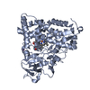
| Deposited unit | 
| ||||||||
|---|---|---|---|---|---|---|---|---|---|
| 1 | 
| ||||||||
| 2 | 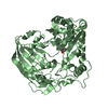
| ||||||||
| Unit cell |
|
-Components
| #1: Protein | Mass: 47177.602 Da / Num. of mol.: 2 Source method: isolated from a genetically manipulated source Source: (gene. exp.) Solenostemon scutellarioides (plant) / Gene: cbhct2 / Production host:  Escherichia coli (E. coli) / Strain (production host): BL21(DE3) Escherichia coli (E. coli) / Strain (production host): BL21(DE3)References: UniProt: E8ZAP2, shikimate O-hydroxycinnamoyltransferase#2: Chemical | 3-Hydroxyacetophenone  Mass: 136.148 Da / Num. of mol.: 2 / Source method: obtained synthetically / Formula: C8H8O2 Mass: 136.148 Da / Num. of mol.: 2 / Source method: obtained synthetically / Formula: C8H8O2#3: Water | ChemComp-HOH / | Water Mass: 18.015 Da / Num. of mol.: 865 / Source method: isolated from a natural source / Formula: H2O Mass: 18.015 Da / Num. of mol.: 865 / Source method: isolated from a natural source / Formula: H2O |
|---|
-Experimental details
-Experiment
| Experiment | Method: X-RAY DIFFRACTION / Number of used crystals: 1 |
|---|
- Sample preparation
| Crystal | Density Matthews: 2.22 Å3/Da / Density % sol: 44.6 % |
|---|---|
| Crystal grow | Temperature: 277.15 K / Method: vapor diffusion, hanging drop / pH: 7.5 Details: 0.1 M Bis-Tris:HCl, 25% PEG 3350, pH 7.5. A soaking drop was prepared with 10% (v/v) of 500 mM 3-hydroxyacetophenone, and 10% (v/v) of 100 mM p-coumaroyl CoA, in reservoir solution. |
-Data collection
| Diffraction | Mean temperature: 100 K |
|---|---|
| Diffraction source | Source: SYNCHROTRON / Site: APS / Beamline: 24-ID-E / Wavelength: 0.979 Å |
| Detector | Type: ADSC QUANTUM 315 / Detector: CCD / Date: Mar 30, 2015 |
| Radiation | Protocol: SINGLE WAVELENGTH / Monochromatic (M) / Laue (L): M / Scattering type: x-ray |
| Radiation wavelength | Wavelength: 0.979 Å / Relative weight: 1 |
| Reflection | Resolution: 1.9→53.54 Å / Num. obs: 95145 / % possible obs: 87.7 % / Redundancy: 2.7 % / Net I/σ(I): 5.9 |
- Processing
| Software |
| ||||||||||||||||||||||||||||||||||||||||||||||||||||||||||||||||||||||||||||||||||||||||||||||||||||||||||||||||||||||||||||||||||||||||||||||||||||||||||||||||||||||||||||||||||||||
|---|---|---|---|---|---|---|---|---|---|---|---|---|---|---|---|---|---|---|---|---|---|---|---|---|---|---|---|---|---|---|---|---|---|---|---|---|---|---|---|---|---|---|---|---|---|---|---|---|---|---|---|---|---|---|---|---|---|---|---|---|---|---|---|---|---|---|---|---|---|---|---|---|---|---|---|---|---|---|---|---|---|---|---|---|---|---|---|---|---|---|---|---|---|---|---|---|---|---|---|---|---|---|---|---|---|---|---|---|---|---|---|---|---|---|---|---|---|---|---|---|---|---|---|---|---|---|---|---|---|---|---|---|---|---|---|---|---|---|---|---|---|---|---|---|---|---|---|---|---|---|---|---|---|---|---|---|---|---|---|---|---|---|---|---|---|---|---|---|---|---|---|---|---|---|---|---|---|---|---|---|---|---|---|
| Refinement | Resolution: 1.9→53.537 Å / SU ML: 0.27 / Cross valid method: FREE R-VALUE / σ(F): 1.96 / Phase error: 27.35 / Stereochemistry target values: ML
| ||||||||||||||||||||||||||||||||||||||||||||||||||||||||||||||||||||||||||||||||||||||||||||||||||||||||||||||||||||||||||||||||||||||||||||||||||||||||||||||||||||||||||||||||||||||
| Solvent computation | Shrinkage radii: 0.9 Å / VDW probe radii: 1.11 Å / Solvent model: FLAT BULK SOLVENT MODEL | ||||||||||||||||||||||||||||||||||||||||||||||||||||||||||||||||||||||||||||||||||||||||||||||||||||||||||||||||||||||||||||||||||||||||||||||||||||||||||||||||||||||||||||||||||||||
| Refinement step | Cycle: LAST / Resolution: 1.9→53.537 Å
| ||||||||||||||||||||||||||||||||||||||||||||||||||||||||||||||||||||||||||||||||||||||||||||||||||||||||||||||||||||||||||||||||||||||||||||||||||||||||||||||||||||||||||||||||||||||
| Refine LS restraints |
| ||||||||||||||||||||||||||||||||||||||||||||||||||||||||||||||||||||||||||||||||||||||||||||||||||||||||||||||||||||||||||||||||||||||||||||||||||||||||||||||||||||||||||||||||||||||
| LS refinement shell |
|
