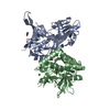
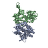
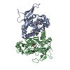
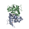
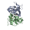
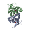
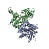
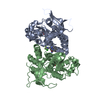
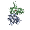
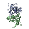
[English] 日本語
Yorodumi
- PDB-5kdt: Structure of the human GluN1/GluN2A LBD in complex with GNE0723 -

+
Open data


ID or keywords:

Loading...

-
Basic information

Entry
Database: PDB / ID: 5kdt
TitleStructure of the human GluN1/GluN2A LBD in complex with GNE0723
Components(Glutamate receptor ionotropic, NMDA ...) x 2
KeywordsTRANSPORT PROTEIN / NMDA / receptor / glutamate / channel
Function / homology
Function and homology information


excitatory chemical synaptic transmission / directional locomotion / Synaptic adhesion-like molecules / serotonin metabolic process / protein localization to postsynaptic membrane / propylene metabolic process / response to glycine / sleep / activation of cysteine-type endopeptidase activity / glutamate-gated calcium ion channel activity ...excitatory chemical synaptic transmission / directional locomotion / Synaptic adhesion-like molecules / serotonin metabolic process / protein localization to postsynaptic membrane / propylene metabolic process / response to glycine / sleep / activation of cysteine-type endopeptidase activity / glutamate-gated calcium ion channel activity / glutamate receptor signaling pathway / regulation of monoatomic cation transmembrane transport / Assembly and cell surface presentation of NMDA receptors / Neurexins and neuroligins / NMDA glutamate receptor activity / NMDA selective glutamate receptor complex / calcium ion transmembrane import into cytosol / protein heterotetramerization / glutamate binding / positive regulation of reactive oxygen species biosynthetic process / glycine binding / positive regulation of calcium ion transport into cytosol / dopamine metabolic process / Negative regulation of NMDA receptor-mediated neuronal transmission / startle response / Unblocking of NMDA receptors, glutamate binding and activation / regulation of neuronal synaptic plasticity / monoatomic cation transmembrane transport / positive regulation of excitatory postsynaptic potential / Long-term potentiation / monoatomic cation transport / ligand-gated monoatomic ion channel activity / excitatory synapse / calcium ion homeostasis / synaptic cleft / MECP2 regulates neuronal receptors and channels / sensory perception of pain / response to amphetamine / EPHB-mediated forward signaling / excitatory postsynaptic potential / regulation of membrane potential / ionotropic glutamate receptor signaling pathway / Ras activation upon Ca2+ influx through NMDA receptor / neurogenesis / positive regulation of synaptic transmission, glutamatergic / transmitter-gated monoatomic ion channel activity involved in regulation of postsynaptic membrane potential / synaptic membrane / synaptic transmission, glutamatergic / long-term synaptic potentiation / postsynaptic density membrane / brain development / protein catabolic process / regulation of synaptic plasticity / visual learning / cytoplasmic vesicle membrane / terminal bouton / negative regulation of protein catabolic process / memory / response to wounding / synaptic vesicle / signaling receptor activity / presynaptic membrane / amyloid-beta binding / chemical synaptic transmission / RAF/MAP kinase cascade / postsynaptic membrane / response to ethanol / dendritic spine / postsynaptic density / learning or memory / calmodulin binding / neuron projection / response to xenobiotic stimulus / positive regulation of apoptotic process / dendrite / synapse / glutamatergic synapse / calcium ion binding / protein-containing complex binding / endoplasmic reticulum membrane / cell surface / positive regulation of transcription by RNA polymerase II / zinc ion binding / plasma membrane / cytoplasm
Similarity search - Function
Glutamate [NMDA] receptor, epsilon subunit, C-terminal / N-methyl D-aspartate receptor 2B3 C-terminus / Bacterial extracellular solute-binding proteins, family 3 / Solute-binding protein family 3/N-terminal domain of MltF / Ionotropic glutamate receptor, metazoa / Ligated ion channel L-glutamate- and glycine-binding site / : / Ligand-gated ion channel / Ionotropic glutamate receptor, L-glutamate and glycine-binding domain / Ligated ion channel L-glutamate- and glycine-binding site ...Glutamate [NMDA] receptor, epsilon subunit, C-terminal / N-methyl D-aspartate receptor 2B3 C-terminus / Bacterial extracellular solute-binding proteins, family 3 / Solute-binding protein family 3/N-terminal domain of MltF / Ionotropic glutamate receptor, metazoa / Ligated ion channel L-glutamate- and glycine-binding site / : / Ligand-gated ion channel / Ionotropic glutamate receptor, L-glutamate and glycine-binding domain / Ligated ion channel L-glutamate- and glycine-binding site / Ionotropic glutamate receptor / Eukaryotic homologues of bacterial periplasmic substrate binding proteins. / Receptor, ligand binding region / Receptor family ligand binding region / Periplasmic binding protein-like II / Periplasmic binding protein-like I / D-Maltodextrin-Binding Protein; domain 2 / 3-Layer(aba) Sandwich / Alpha Beta
Similarity search - Domain/homology
Chem-6RV / ACETATE ION / GLUTAMIC ACID / GLYCINE / Glutamate receptor ionotropic, NMDA 1 / Glutamate receptor ionotropic, NMDA 2A
Similarity search - Component
Biological speciesHomo sapiens (human)
MethodX-RAY DIFFRACTION / SYNCHROTRON / MOLECULAR REPLACEMENT / Resolution: 2.44 Å
AuthorsWallweber, H.J.A. / Lupardus, P.J.
CitationJournal: J. Med. Chem. / Year: 2016
Title: Discovery of GluN2A-Selective NMDA Receptor Positive Allosteric Modulators (PAMs): Tuning Deactivation Kinetics via Structure-Based Design.
Authors: Volgraf, M. / Sellers, B.D. / Jiang, Y. / Wu, G. / Ly, C.Q. / Villemure, E. / Pastor, R.M. / Yuen, P.W. / Lu, A. / Luo, X. / Liu, M. / Zhang, S. / Sun, L. / Fu, Y. / Lupardus, P.J. / ...Authors: Volgraf, M. / Sellers, B.D. / Jiang, Y. / Wu, G. / Ly, C.Q. / Villemure, E. / Pastor, R.M. / Yuen, P.W. / Lu, A. / Luo, X. / Liu, M. / Zhang, S. / Sun, L. / Fu, Y. / Lupardus, P.J. / Wallweber, H.J. / Liederer, B.M. / Deshmukh, G. / Plise, E. / Tay, S. / Reynen, P. / Herrington, J. / Gustafson, A. / Liu, Y. / Dirksen, A. / Dietz, M.G. / Liu, Y. / Wang, T.M. / Hanson, J.E. / Hackos, D. / Scearce-Levie, K. / Schwarz, J.B.
History
DepositionJun 8, 2016Deposition site: RCSB / Processing site: RCSB
SupersessionJul 13, 2016ID: 5I2J
Revision 1.0Jul 13, 2016Provider: repository / Type: Initial release

-
Structure visualization

Structure viewerMolecule:
MolmilJmol/JSmol

Downloads & links

-
Assembly

Deposited unit
A: Glutamate receptor ionotropic, NMDA 2A
B: Glutamate receptor ionotropic, NMDA 1
hetero molecules


Theoretical massNumber of molelcules
Total (without water)66,0426
Polymers65,2932
Non-polymers7494
Water2,774154
1


  • Idetical with deposited unit
  • defined by author&software
TypeNameSymmetry operationNumber
identity operation1_555x,y,z1
Buried area2450 Å2
ΔGint-4 kcal/mol
Surface area25300 Å2
MethodPISA
Unit cell
Length a, b, c (Å)55.325, 90.127, 123.185
Angle α, β, γ (deg.)90.00, 90.00, 90.00
Int Tables number19
Space group name H-MP212121

-
Components

-
Glutamate receptor ionotropic, NMDA ... , 2 types, 2 molecules AB

#1: Protein Glutamate receptor ionotropic, NMDA 2A / GluN2A / Glutamate [NMDA] receptor subunit epsilon-1 / N-methyl D-aspartate receptor subtype 2A / hNR2A


Mass: 31883.441 Da / Num. of mol.: 1
Fragment: Ligand binding domain (UNP residues 401-539, GT linker, UNP residues 661-802)
Source method: isolated from a genetically manipulated source
Source: (gene. exp.) Homo sapiens (human) / Gene: GRIN2A, NMDAR2A / Production host: Escherichia coli (E. coli) / References: UniProt: Q12879
#2: Protein Glutamate receptor ionotropic, NMDA 1 / GluN1 / Glutamate [NMDA] receptor subunit zeta-1 / N-methyl-D-aspartate receptor subunit NR1 / NMD-R1


Mass: 33409.074 Da / Num. of mol.: 1
Fragment: Ligand binding domain (UNP residues 415-565, GT linker, UNP residues 684-821)
Source method: isolated from a genetically manipulated source
Source: (gene. exp.) Homo sapiens (human) / Gene: GRIN1, NMDAR1 / Production host: Escherichia coli (E. coli) / References: UniProt: Q05586

-
Non-polymers , 5 types, 158 molecules

#3: Chemical ChemComp-ACT / ACETATE ION / Acetate


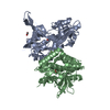
Mass: 59.044 Da / Num. of mol.: 1
Source method: isolated from a genetically manipulated source
Formula: C2H3O2
#4: Chemical ChemComp-GLU / GLUTAMIC ACID / Glutamic acid


Type: L-peptide linking / Mass: 147.129 Da / Num. of mol.: 1 / Source method: obtained synthetically / Formula: C5H9NO4
#5: Chemical ChemComp-6RV / (1~{R},2~{R})-2-[7-[[5-chloranyl-3-(trifluoromethyl)pyrazol-1-yl]methyl]-5-oxidanylidene-2-(trifluoromethyl)-[1,3]thiazolo[3,2-a]pyrimidin-3-yl]cyclopropane-1-carbonitrile


Mass: 467.776 Da / Num. of mol.: 1 / Source method: obtained synthetically / Formula: C16H8ClF6N5OS
#6: Chemical ChemComp-GLY / GLYCINE / Glycine


Type: peptide linking / Mass: 75.067 Da / Num. of mol.: 1
Source method: isolated from a genetically manipulated source
Formula: C2H5NO2
#7: Water ChemComp-HOH / water / Water


Mass: 18.015 Da / Num. of mol.: 154 / Source method: isolated from a natural source / Formula: H2O

-
Experimental details

-
Experiment

ExperimentMethod: X-RAY DIFFRACTION / Number of used crystals: 1

-
Sample preparation

CrystalDensity Matthews: 2.45 Å3/Da / Density % sol: 49.78 %
Crystal growTemperature: 277 K / Method: vapor diffusion, hanging drop / pH: 7
Details: 0.1 M HEPES, pH 7.0, 10-13% PEG8000, 2 mM calcium acetate

-
Data collection

DiffractionMean temperature: 100 K
Diffraction sourceSource: SYNCHROTRON / Site: ALS / Beamline: 5.0.2 / Wavelength: 1 Å
DetectorType: ADSC QUANTUM 315r / Detector: CCD / Date: Sep 10, 2014
RadiationMonochromator: double crystal Si(111) / Protocol: SINGLE WAVELENGTH / Monochromatic (M) / Laue (L): M / Scattering type: x-ray
Radiation wavelengthWavelength: 1 Å / Relative weight: 1
ReflectionResolution: 2.44→90 Å / Num. obs: 23645 / % possible obs: 100 % / Redundancy: 7.1 % / Biso Wilson estimate: 62.31 Å2 / Rmerge(I) obs: 0.075 / Net I/σ(I): 18.1
Reflection shellResolution: 2.44→2.57 Å / Redundancy: 7.3 % / Rmerge(I) obs: 0.802 / Mean I/σ(I) obs: 2.4 / % possible all: 100

-
Processing

Software
NameVersionClassification
BUSTER2.11.5refinement
XDSdata reduction
XSCALEdata scaling
PHASERphasing
RefinementMethod to determine structure: MOLECULAR REPLACEMENT / Resolution: 2.44→72.74 Å / Cor.coef. Fo:Fc: 0.9448 / Cor.coef. Fo:Fc free: 0.9203 / SU R Cruickshank DPI: 0.425 / Cross valid method: THROUGHOUT / σ(F): 0 / SU R Blow DPI: 0.458 / SU Rfree Blow DPI: 0.259 / SU Rfree Cruickshank DPI: 0.258
RfactorNum. reflection% reflectionSelection details
Rfree0.2405 1192 5.04 %RANDOM
Rwork0.1861 ---
obs0.1888 23645 99.99 %-
Displacement parametersBiso mean: 57.44 Å2
Baniso -1Baniso -2Baniso -3
1--4.1682 Å20 Å20 Å2
2--6.8969 Å20 Å2
3----2.7287 Å2
Refine analyzeLuzzati coordinate error obs: 0.284 Å
Refinement stepCycle: LAST / Resolution: 2.44→72.74 Å
ProteinNucleic acidLigandSolventTotal
Num. atoms4419 0 49 154 4622
Refine LS restraints
Refine-IDTypeDev idealNumberRestraint functionWeight
X-RAY DIFFRACTIONt_bond_d0.014551HARMONIC2
X-RAY DIFFRACTIONt_angle_deg1.136163HARMONIC2
X-RAY DIFFRACTIONt_dihedral_angle_d1575SINUSOIDAL2
X-RAY DIFFRACTIONt_incorr_chiral_ct
X-RAY DIFFRACTIONt_pseud_angle
X-RAY DIFFRACTIONt_trig_c_planes107HARMONIC2
X-RAY DIFFRACTIONt_gen_planes677HARMONIC5
X-RAY DIFFRACTIONt_it4551HARMONIC20
X-RAY DIFFRACTIONt_nbd
X-RAY DIFFRACTIONt_omega_torsion3.02
X-RAY DIFFRACTIONt_other_torsion19.27
X-RAY DIFFRACTIONt_improper_torsion
X-RAY DIFFRACTIONt_chiral_improper_torsion596SEMIHARMONIC5
X-RAY DIFFRACTIONt_sum_occupancies
X-RAY DIFFRACTIONt_utility_distance
X-RAY DIFFRACTIONt_utility_angle
X-RAY DIFFRACTIONt_utility_torsion
X-RAY DIFFRACTIONt_ideal_dist_contact5231SEMIHARMONIC4
LS refinement shellResolution: 2.44→2.55 Å / Total num. of bins used: 12
RfactorNum. reflection% reflection
Rfree0.2919 134 4.68 %
Rwork0.2185 2732 -
all0.2221 2866 -
obs--99.99 %

+
About Yorodumi

-
News

-
Feb 9, 2022. New format data for meta-information of EMDB entries

New format data for meta-information of EMDB entries

  • Version 3 of the EMDB header file is now the official format.
  • The previous official version 1.9 will be removed from the archive.

Related info.:EMDB header

External links:wwPDB to switch to version 3 of the EMDB data model

-
Aug 12, 2020. Covid-19 info

Covid-19 info

URL: https://pdbj.org/emnavi/covid19.php

New page: Covid-19 featured information page in EM Navigator.

Related info.:Covid-19 info / Mar 5, 2020. Novel coronavirus structure data

+
Mar 5, 2020. Novel coronavirus structure data

Novel coronavirus structure data

Related info.:Yorodumi Speices / Aug 12, 2020. Covid-19 info

External links:COVID-19 featured content - PDBj / Molecule of the Month (242):Coronavirus Proteases

+
Jan 31, 2019. EMDB accession codes are about to change! (news from PDBe EMDB page)

EMDB accession codes are about to change! (news from PDBe EMDB page)

  • The allocation of 4 digits for EMDB accession codes will soon come to an end. Whilst these codes will remain in use, new EMDB accession codes will include an additional digit and will expand incrementally as the available range of codes is exhausted. The current 4-digit format prefixed with “EMD-” (i.e. EMD-XXXX) will advance to a 5-digit format (i.e. EMD-XXXXX), and so on. It is currently estimated that the 4-digit codes will be depleted around Spring 2019, at which point the 5-digit format will come into force.
  • The EM Navigator/Yorodumi systems omit the EMD- prefix.

Related info.:Q: What is EMD? / ID/Accession-code notation in Yorodumi/EM Navigator

External links:EMDB Accession Codes are Changing Soon! / Contact to PDBj

+
Jul 12, 2017. Major update of PDB

Major update of PDB

  • wwPDB released updated PDB data conforming to the new PDBx/mmCIF dictionary.
  • This is a major update changing the version number from 4 to 5, and with Remediation, in which all the entries are updated.
  • In this update, many items about electron microscopy experimental information are reorganized (e.g. em_software).
  • Now, EM Navigator and Yorodumi are based on the updated data.

External links:wwPDB Remediation / Enriched Model Files Conforming to OneDep Data Standards Now Available in the PDB FTP Archive

-
Yorodumi

Thousand views of thousand structures

  • Yorodumi is a browser for structure data from EMDB, PDB, SASBDB, etc.
  • This page is also the successor to EM Navigator detail page, and also detail information page/front-end page for Omokage search.
  • The word "yorodu" (or yorozu) is an old Japanese word meaning "ten thousand". "mi" (miru) is to see.

Related info.:EMDB / PDB / SASBDB / Comparison of 3 databanks / Yorodumi Search / Aug 31, 2016. New EM Navigator & Yorodumi / Yorodumi Papers / Jmol/JSmol / Function and homology information / Changes in new EM Navigator and Yorodumi

Read more