 Movie
Movie Controller

[English] 日本語
 Yorodumi
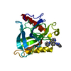
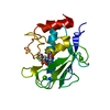
- PDB-5k9y: Crystal structure of a thermophilic xylanase A from Bacillus subt... -
+ Open data

- Basic information
| Entry | Database: PDB / ID: 5k9y | |||||||||
|---|---|---|---|---|---|---|---|---|---|---|
| Title | Crystal structure of a thermophilic xylanase A from Bacillus subtilis 1A1 quadruple mutant Q7H/G13R/S22P/S179C | |||||||||
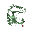
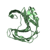
 Components Components | Endo-1,4-beta-xylanase A | |||||||||
 Keywords Keywords |  HYDROLASE / Glycoside Hydrolase Family 11 / Endo-1 / 4-beta-xylanase A / thermostability HYDROLASE / Glycoside Hydrolase Family 11 / Endo-1 / 4-beta-xylanase A / thermostability | |||||||||
| Function / homology |  Function and homology informationendo-1,4-beta-xylanase activity / endo-1,4-beta-xylanase / xylan catabolic process Function and homology informationendo-1,4-beta-xylanase activity / endo-1,4-beta-xylanase / xylan catabolic processSimilarity search - Function | |||||||||
| Biological species |  Bacillus subtilis (bacteria) Bacillus subtilis (bacteria) | |||||||||
| Method | X-RAY DIFFRACTION / SYNCHROTRON / MOLECULAR REPLACEMENT / molecular replacement / Resolution: 2.2 Å | |||||||||
 Authors Authors | Pinheiro, M.P. / Ferreira, T.L. / Silva, S.R.B. / Fuzo, C.A. / Silva, S.R. / Lourenzoni, M.R. / Vieira, D.S. / Ward, R.J. / Nonato, M.C. | |||||||||
| Funding support |  Brazil, 2items Brazil, 2items
| |||||||||
 Citation Citation | Journal: Int. J. Biol. Macromol. / Year: 2017 Title: The role of local residue environmental changes in thermostable mutants of the GH11 xylanase from Bacillus subtilis. Authors: Silva, S.B. / Pinheiro, M.P. / Fuzo, C.A. / Silva, S.R. / Ferreira, T.L. / Lourenzoni, M.R. / Nonato, M.C. / Vieira, D.S. / Ward, R.J. | |||||||||
| History |
|
- Structure visualization
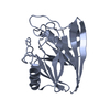
| Structure viewer | Molecule: MolmilJmol/JSmol |
|---|
- Downloads & links
-Download
| PDBx/mmCIF format | 5k9y.cif.gz | 90.4 KB | Display | PDBx/mmCIF format |
|---|---|---|---|---|
| PDB format | pdb5k9y.ent.gz | 67.6 KB | Display | PDB format |
| PDBx/mmJSON format | 5k9y.json.gz | Tree view | PDBx/mmJSON format | |
| Others |  Other downloads Other downloads |
-Validation report
| Arichive directory | https://data.pdbj.org/pub/pdb/validation_reports/k9/5k9yftp://data.pdbj.org/pub/pdb/validation_reports/k9/5k9y | HTTPS FTP |
|---|
-Related structure data
| Related structure data |  1xxnS S: Starting model for refinement |
|---|---|
| Similar structure data |


-Links
 PDBj
PDBj
- Assembly
| Deposited unit | 
| ||||||||
|---|---|---|---|---|---|---|---|---|---|
| 1 | 
| ||||||||
| 2 | 
| ||||||||
| Unit cell |
| ||||||||
| Details | Monomer as determined by gel filtration. |
-Components
| #1: Protein | Mass: 20547.240 Da / Num. of mol.: 2 / Fragment: residues 29-213 / Mutation: Q7H, G13R, S22P, S179C Source method: isolated from a genetically manipulated source Source: (gene. exp.) Bacillus subtilis (strain 168) (bacteria)Strain: 168 / Gene: xynA, BSU18840 / Production host: Escherichia coli (E. coli) / References: UniProt: P18429, endo-1,4-beta-xylanase#2: Water | ChemComp-HOH / | Water Mass: 18.015 Da / Num. of mol.: 209 / Source method: isolated from a natural source / Formula: H2O Mass: 18.015 Da / Num. of mol.: 209 / Source method: isolated from a natural source / Formula: H2O |
|---|
-Experimental details
-Experiment
| Experiment | Method: X-RAY DIFFRACTION / Number of used crystals: 1 |
|---|
- Sample preparation
| Crystal | Density Matthews: 2.03 Å3/Da / Density % sol: 39.27 % |
|---|---|
| Crystal grow | Temperature: 295 K / Method: vapor diffusion, sitting drop / pH: 7 / Details: 0.1 M HEPES and 0.6 M sodium tartrate |
-Data collection
| Diffraction | Mean temperature: 100 K | ||||||||||||||||||||||||||||||||||||||||||||||||||||||||||||||||||
|---|---|---|---|---|---|---|---|---|---|---|---|---|---|---|---|---|---|---|---|---|---|---|---|---|---|---|---|---|---|---|---|---|---|---|---|---|---|---|---|---|---|---|---|---|---|---|---|---|---|---|---|---|---|---|---|---|---|---|---|---|---|---|---|---|---|---|---|
| Diffraction source | Source: SYNCHROTRON / Site: LNLS / Beamline: W01B-MX2 / Wavelength: 1.5497 Å | ||||||||||||||||||||||||||||||||||||||||||||||||||||||||||||||||||
| Detector | Type: MARMOSAIC 225 mm CCD / Detector: CCD / Date: Jul 12, 2007 | ||||||||||||||||||||||||||||||||||||||||||||||||||||||||||||||||||
| Radiation | Monochromator: Si(111) double-crystal / Protocol: SINGLE WAVELENGTH / Monochromatic (M) / Laue (L): M / Scattering type: x-ray | ||||||||||||||||||||||||||||||||||||||||||||||||||||||||||||||||||
| Radiation wavelength | Wavelength: 1.5497 Å / Relative weight: 1 | ||||||||||||||||||||||||||||||||||||||||||||||||||||||||||||||||||
| Reflection | Resolution: 2.2→42.247 Å / Num. obs: 16886 / % possible obs: 100 % / Redundancy: 3.6 % / Rsym value: 0.09 / Net I/av σ(I): 5.41 / Net I/σ(I): 9.1 | ||||||||||||||||||||||||||||||||||||||||||||||||||||||||||||||||||
| Reflection shell |
|
-Phasing
| Phasing | Method: molecular replacement |
|---|
- Processing
| Software |
| |||||||||||||||||||||||||||||||||||||||||||||
|---|---|---|---|---|---|---|---|---|---|---|---|---|---|---|---|---|---|---|---|---|---|---|---|---|---|---|---|---|---|---|---|---|---|---|---|---|---|---|---|---|---|---|---|---|---|---|
| Refinement | Method to determine structure: MOLECULAR REPLACEMENT Starting model: 1XXN Resolution: 2.2→20.33 Å / Cor.coef. Fo:Fc: 0.945 / Cor.coef. Fo:Fc free: 0.899 / SU R Cruickshank DPI: 0.4036 / Cross valid method: THROUGHOUT / σ(F): 0 / ESU R: 0.404 / ESU R Free: 0.248 Details: HYDROGENS HAVE BEEN USED IF PRESENT IN THE INPUT U VALUES : REFINED INDIVIDUALLY
| |||||||||||||||||||||||||||||||||||||||||||||
| Solvent computation | Ion probe radii: 0.8 Å / Shrinkage radii: 0.8 Å / VDW probe radii: 1.2 Å | |||||||||||||||||||||||||||||||||||||||||||||
| Displacement parameters | Biso max: 68.2 Å2 / Biso mean: 26.818 Å2 / Biso min: 9.47 Å2
| |||||||||||||||||||||||||||||||||||||||||||||
| Refinement step | Cycle: final / Resolution: 2.2→20.33 Å
| |||||||||||||||||||||||||||||||||||||||||||||
| Refine LS restraints |
| |||||||||||||||||||||||||||||||||||||||||||||
| LS refinement shell | Resolution: 2.2→2.257 Å / Total num. of bins used: 20
|
