 Movie
Movie Controller

[English] 日本語
 Yorodumi
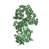
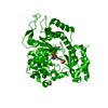
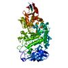
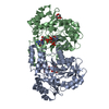
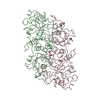
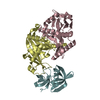
- PDB-5k8b: X-ray structure of KdnA, 8-amino-3,8-dideoxy-alpha-D-manno-octulo... -
+ Open data

- Basic information
| Entry | Database: PDB / ID: 5k8b | ||||||
|---|---|---|---|---|---|---|---|
| Title | X-ray structure of KdnA, 8-amino-3,8-dideoxy-alpha-D-manno-octulosonate transaminase, from Shewanella oneidensis in the presence of the external aldimine with PLP and glutamate | ||||||
 Components Components | 8-amino-3,8-dideoxy-alpha-D-manno-octulosonate transaminase | ||||||
 Keywords Keywords |  TRANSFERASE / Transaminase / 8-amino-3 / 8-dideoxy-d-manno-octulosonic acid / deoxy sugar TRANSFERASE / Transaminase / 8-amino-3 / 8-dideoxy-d-manno-octulosonic acid / deoxy sugar | ||||||
| Function / homology |  Function and homology information Function and homology information8-amino-3,8-dideoxy-alpha-D-manno-octulosonate transaminase / polysaccharide biosynthetic process / lipopolysaccharide biosynthetic process / transaminase activity / catalytic activity / pyridoxal phosphate bindingSimilarity search - Function | ||||||
| Biological species |  Shewanella oneidensis (bacteria) Shewanella oneidensis (bacteria) | ||||||
| Method | X-RAY DIFFRACTION / MOLECULAR REPLACEMENT / Resolution: 2.15 Å | ||||||
 Authors Authors | Holden, H.M. / Thoden, J.B. / Zachman-Brockmeyer, T.R. | ||||||
| Funding support |  United States, 1items United States, 1items
| ||||||
 Citation Citation | Journal: Biochemistry / Year: 2016 Title: Structures of KdnB and KdnA from Shewanella oneidensis: Key Enzymes in the Formation of 8-Amino-3,8-Dideoxy-d-Manno-Octulosonic Acid. Authors: Zachman-Brockmeyer, T.R. / Thoden, J.B. / Holden, H.M. | ||||||
| History |
|
- Structure visualization
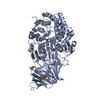
| Structure viewer | Molecule: MolmilJmol/JSmol |
|---|
- Downloads & links
-Download
| PDBx/mmCIF format | 5k8b.cif.gz | 325.2 KB | Display | PDBx/mmCIF format |
|---|---|---|---|---|
| PDB format | pdb5k8b.ent.gz | 263.7 KB | Display | PDB format |
| PDBx/mmJSON format | 5k8b.json.gz | Tree view | PDBx/mmJSON format | |
| Others |  Other downloads Other downloads |
-Validation report
| Arichive directory | https://data.pdbj.org/pub/pdb/validation_reports/k8/5k8bftp://data.pdbj.org/pub/pdb/validation_reports/k8/5k8b | HTTPS FTP |
|---|
-Related structure data
| Related structure data | 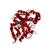 5k8cC  3frkS C: citing same article ( S: Starting model for refinement |
|---|---|
| Similar structure data |

-Links
 PDBj
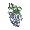
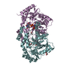
PDBj- Assembly
| Deposited unit | 
| ||||||||
|---|---|---|---|---|---|---|---|---|---|
| 1 | 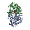
| ||||||||
| 2 | 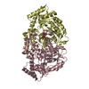
| ||||||||
| Unit cell |
|
-Components
| #1: Protein | Mass: 44768.273 Da / Num. of mol.: 4 Source method: isolated from a genetically manipulated source Source: (gene. exp.) Shewanella oneidensis (strain MR-1) (bacteria)Strain: MR-1 / Gene: kdnA, SO_2476 / Production host: Escherichia coli (E. coli)References: UniProt: Q8EEB1, 8-amino-3,8-dideoxy-alpha-D-manno-octulosonate transaminase #2: Chemical | ChemComp-PDG /   Mass: 378.272 Da / Num. of mol.: 4 / Source method: obtained synthetically / Formula: C13H19N2O9P Mass: 378.272 Da / Num. of mol.: 4 / Source method: obtained synthetically / Formula: C13H19N2O9P#3: Chemical | ChemComp-CL / Chloride  Mass: 35.453 Da / Num. of mol.: 5 / Source method: obtained synthetically / Formula: Cl Mass: 35.453 Da / Num. of mol.: 5 / Source method: obtained synthetically / Formula: Cl#4: Chemical | ChemComp-NA / |   Mass: 22.990 Da / Num. of mol.: 1 / Source method: obtained synthetically / Formula: Na Mass: 22.990 Da / Num. of mol.: 1 / Source method: obtained synthetically / Formula: Na#5: Water | ChemComp-HOH / | Water Mass: 18.015 Da / Num. of mol.: 710 / Source method: isolated from a natural source / Formula: H2O Mass: 18.015 Da / Num. of mol.: 710 / Source method: isolated from a natural source / Formula: H2O |
|---|
-Experimental details
-Experiment
| Experiment | Method: X-RAY DIFFRACTION / Number of used crystals: 1 |
|---|
- Sample preparation
| Crystal | Density Matthews: 2.24 Å3/Da / Density % sol: 45.02 % |
|---|---|
| Crystal grow | Temperature: 295 K / Method: vapor diffusion, hanging drop / pH: 6 Details: 16-21% PEG-3350, 100 mM MES, 200 mM KCl, 150 mM MgCl2, 1 mM PLP, 50 mM monosodium glutamate |
-Data collection
| Diffraction | Mean temperature: 100 K |
|---|---|
| Diffraction source | Source: ROTATING ANODE / Type: RIGAKU RU200 / Wavelength: 1.5418 Å |
| Detector | Type: Bruker Platinum 135 / Detector: CCD / Date: Sep 22, 2015 |
| Radiation | Protocol: SINGLE WAVELENGTH / Monochromatic (M) / Laue (L): M / Scattering type: x-ray |
| Radiation wavelength | Wavelength: 1.5418 Å / Relative weight: 1 |
| Reflection | Resolution: 2.15→50 Å / Num. obs: 83041 / % possible obs: 95.8 % / Observed criterion σ(F): 0 / Observed criterion σ(I): 0 / Redundancy: 2.9 % / Rmerge(I) obs: 0.078 / Rsym value: 0.078 / Net I/σ(I): 7.8 |
| Reflection shell | Resolution: 2.15→2.25 Å / Redundancy: 1.8 % / Rmerge(I) obs: 0.254 / Mean I/σ(I) obs: 2.4 / % possible all: 89.4 |
- Processing
| Software |
| ||||||||||||||||||||||||||||||||||||||||||||||||||||||||||||||||||||||||||||||||||||||||||||||||||||||||||||||||||||||||||||||||||||||||||||||||||||||||||||||||||||||||||||||||||||||
|---|---|---|---|---|---|---|---|---|---|---|---|---|---|---|---|---|---|---|---|---|---|---|---|---|---|---|---|---|---|---|---|---|---|---|---|---|---|---|---|---|---|---|---|---|---|---|---|---|---|---|---|---|---|---|---|---|---|---|---|---|---|---|---|---|---|---|---|---|---|---|---|---|---|---|---|---|---|---|---|---|---|---|---|---|---|---|---|---|---|---|---|---|---|---|---|---|---|---|---|---|---|---|---|---|---|---|---|---|---|---|---|---|---|---|---|---|---|---|---|---|---|---|---|---|---|---|---|---|---|---|---|---|---|---|---|---|---|---|---|---|---|---|---|---|---|---|---|---|---|---|---|---|---|---|---|---|---|---|---|---|---|---|---|---|---|---|---|---|---|---|---|---|---|---|---|---|---|---|---|---|---|---|---|
| Refinement | Method to determine structure: MOLECULAR REPLACEMENT Starting model: PDB entry 3FRK Resolution: 2.15→50 Å / Cor.coef. Fo:Fc: 0.951 / Cor.coef. Fo:Fc free: 0.916 / SU B: 6.549 / SU ML: 0.163 / Cross valid method: THROUGHOUT / ESU R: 0.265 / ESU R Free: 0.203 / Details: HYDROGENS HAVE BEEN ADDED IN THE RIDING POSITIONS
| ||||||||||||||||||||||||||||||||||||||||||||||||||||||||||||||||||||||||||||||||||||||||||||||||||||||||||||||||||||||||||||||||||||||||||||||||||||||||||||||||||||||||||||||||||||||
| Solvent computation | Ion probe radii: 0.8 Å / Shrinkage radii: 0.8 Å / VDW probe radii: 1.2 Å | ||||||||||||||||||||||||||||||||||||||||||||||||||||||||||||||||||||||||||||||||||||||||||||||||||||||||||||||||||||||||||||||||||||||||||||||||||||||||||||||||||||||||||||||||||||||
| Displacement parameters | Biso mean: 22.988 Å2
| ||||||||||||||||||||||||||||||||||||||||||||||||||||||||||||||||||||||||||||||||||||||||||||||||||||||||||||||||||||||||||||||||||||||||||||||||||||||||||||||||||||||||||||||||||||||
| Refinement step | Cycle: 1 / Resolution: 2.15→50 Å
| ||||||||||||||||||||||||||||||||||||||||||||||||||||||||||||||||||||||||||||||||||||||||||||||||||||||||||||||||||||||||||||||||||||||||||||||||||||||||||||||||||||||||||||||||||||||
| Refine LS restraints |
|