 Movie
Movie Controller

[English] 日本語
 Yorodumi
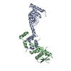
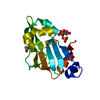
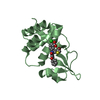
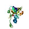
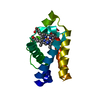
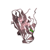
- PDB-5k34: Structure of the ankyrin domain of AnkB from Legionella Pneumophila -
+ Open data

- Basic information
| Entry | Database: PDB / ID: 5k34 | ||||||
|---|---|---|---|---|---|---|---|
| Title | Structure of the ankyrin domain of AnkB from Legionella Pneumophila | ||||||
 Components Components | Ankyrin-repeat protein B | ||||||
 Keywords Keywords |  PROTEIN BINDING / bacterial effector / ankyrin domain / protein-protein interaction / Structural Genomics / Montreal-Kingston Bacterial Structural Genomics Initiative / BSGI PROTEIN BINDING / bacterial effector / ankyrin domain / protein-protein interaction / Structural Genomics / Montreal-Kingston Bacterial Structural Genomics Initiative / BSGI | ||||||
| Function / homology | F-box domain profile. / F-box-like domain superfamily / F-box domain / Ankyrin repeat-containing domain superfamily / Ankyrin-repeat protein B / F-box protein Function and homology information Function and homology information | ||||||
| Biological species |   Legionella pneumophila (bacteria) Legionella pneumophila (bacteria) | ||||||
| Method | X-RAY DIFFRACTION / SYNCHROTRON / SAD / Resolution: 1.15 Å | ||||||
 Authors Authors | Wong, K. / Kozlov, G. / Gehring, K. / Montreal-Kingston Bacterial Structural Genomics Initiative (BSGI) | ||||||
| Funding support |  Canada, 1items Canada, 1items
| ||||||
 Citation Citation | Journal: Structure / Year: 2017 Title: Structural Mimicry by a Bacterial F Box Effector Hijacks the Host Ubiquitin-Proteasome System. Authors: Wong, K. / Perpich, J.D. / Kozlov, G. / Cygler, M. / Abu Kwaik, Y. / Gehring, K. | ||||||
| History |
|
- Structure visualization
| Structure viewer | Molecule: MolmilJmol/JSmol |
|---|
- Downloads & links
-Download
| PDBx/mmCIF format | 5k34.cif.gz | 67.1 KB | Display | PDBx/mmCIF format |
|---|---|---|---|---|
| PDB format | pdb5k34.ent.gz | 48.7 KB | Display | PDB format |
| PDBx/mmJSON format | 5k34.json.gz | Tree view | PDBx/mmJSON format | |
| Others |  Other downloads Other downloads |
-Validation report
| Arichive directory | https://data.pdbj.org/pub/pdb/validation_reports/k3/5k34ftp://data.pdbj.org/pub/pdb/validation_reports/k3/5k34 | HTTPS FTP |
|---|
-Related structure data


-Links
 PDBj
PDBj


- Assembly
| Deposited unit | 
| ||||||||||||
|---|---|---|---|---|---|---|---|---|---|---|---|---|---|
| 1 |
| ||||||||||||
| Unit cell |
| ||||||||||||
| Components on special symmetry positions |
|
-Components
| #1: Protein | Mass: 13768.890 Da / Num. of mol.: 1 Source method: isolated from a genetically manipulated source Source: (gene. exp.) Legionella pneumophila (bacteria) / Gene: ankB / Production host: Escherichia coli (E. coli) / Strain (production host): BL21 / References: UniProt: A0A0A1EKG3, UniProt: Q5ZTL7*PLUS |
|---|---|
| #2: Chemical | ChemComp-GOL / Glycerol  Mass: 92.094 Da / Num. of mol.: 1 / Source method: obtained synthetically / Formula: C3H8O3 Mass: 92.094 Da / Num. of mol.: 1 / Source method: obtained synthetically / Formula: C3H8O3 |
| #3: Chemical | ChemComp-SO4 / Sulfate  Mass: 96.063 Da / Num. of mol.: 1 / Source method: obtained synthetically / Formula: SO4 Mass: 96.063 Da / Num. of mol.: 1 / Source method: obtained synthetically / Formula: SO4 |
| #4: Water | ChemComp-HOH / Water Mass: 18.015 Da / Num. of mol.: 166 / Source method: isolated from a natural source / Formula: H2O Mass: 18.015 Da / Num. of mol.: 166 / Source method: isolated from a natural source / Formula: H2O |
-Experimental details
-Experiment
| Experiment | Method: X-RAY DIFFRACTION / Number of used crystals: 1 |
|---|
- Sample preparation
| Crystal | Density Matthews: 2.15 Å3/Da / Density % sol: 42.7 % |
|---|---|
| Crystal grow | Temperature: 293 K / Method: vapor diffusion, hanging drop Details: 0.2 M lithium sulfate, 0.1 M HEPES pH 7.5, 25% (w/v) PEG 3350 |
-Data collection
| Diffraction | Mean temperature: 100 K |
|---|---|
| Diffraction source | Source: SYNCHROTRON / Site: CHESS  / Beamline: F1 / Wavelength: 0.9179 Å / Beamline: F1 / Wavelength: 0.9179 Å |
| Detector | Type: ADSC QUANTUM 270 / Detector: CCD / Date: Jun 5, 2014 |
| Radiation | Protocol: SINGLE WAVELENGTH / Monochromatic (M) / Laue (L): M / Scattering type: x-ray |
| Radiation wavelength | Wavelength: 0.9179 Å / Relative weight: 1 |
| Reflection | Resolution: 1.15→50 Å / Num. obs: 39731 / % possible obs: 98.6 % / Redundancy: 3.6 % / Rsym value: 0.104 / Net I/σ(I): 22.3 |
| Reflection shell | Resolution: 1.15→1.17 Å / Redundancy: 3.6 % / Rmerge(I) obs: 0.435 / Mean I/σ(I) obs: 3.8 / % possible all: 97.9 |
- Processing
| Software |
| ||||||||||||||||||||||||||||||||||||||||||||||||||||||||||||||||||||||||||||||||||||||||||||||||||||||||||||||||||||||||||||||||||||||||||||||||||||||||||||||||||||||||||||||||||||||
|---|---|---|---|---|---|---|---|---|---|---|---|---|---|---|---|---|---|---|---|---|---|---|---|---|---|---|---|---|---|---|---|---|---|---|---|---|---|---|---|---|---|---|---|---|---|---|---|---|---|---|---|---|---|---|---|---|---|---|---|---|---|---|---|---|---|---|---|---|---|---|---|---|---|---|---|---|---|---|---|---|---|---|---|---|---|---|---|---|---|---|---|---|---|---|---|---|---|---|---|---|---|---|---|---|---|---|---|---|---|---|---|---|---|---|---|---|---|---|---|---|---|---|---|---|---|---|---|---|---|---|---|---|---|---|---|---|---|---|---|---|---|---|---|---|---|---|---|---|---|---|---|---|---|---|---|---|---|---|---|---|---|---|---|---|---|---|---|---|---|---|---|---|---|---|---|---|---|---|---|---|---|---|---|
| Refinement | Method to determine structure: SAD / Resolution: 1.15→45.03 Å / Cor.coef. Fo:Fc: 0.965 / Cor.coef. Fo:Fc free: 0.957 / SU B: 1.264 / SU ML: 0.026 / Cross valid method: THROUGHOUT / ESU R: 0.039 / ESU R Free: 0.038
| ||||||||||||||||||||||||||||||||||||||||||||||||||||||||||||||||||||||||||||||||||||||||||||||||||||||||||||||||||||||||||||||||||||||||||||||||||||||||||||||||||||||||||||||||||||||
| Solvent computation | Ion probe radii: 0.8 Å / Shrinkage radii: 0.8 Å / VDW probe radii: 1.2 Å | ||||||||||||||||||||||||||||||||||||||||||||||||||||||||||||||||||||||||||||||||||||||||||||||||||||||||||||||||||||||||||||||||||||||||||||||||||||||||||||||||||||||||||||||||||||||
| Displacement parameters | Biso mean: 14.613 Å2
| ||||||||||||||||||||||||||||||||||||||||||||||||||||||||||||||||||||||||||||||||||||||||||||||||||||||||||||||||||||||||||||||||||||||||||||||||||||||||||||||||||||||||||||||||||||||
| Refinement step | Cycle: 1 / Resolution: 1.15→45.03 Å
| ||||||||||||||||||||||||||||||||||||||||||||||||||||||||||||||||||||||||||||||||||||||||||||||||||||||||||||||||||||||||||||||||||||||||||||||||||||||||||||||||||||||||||||||||||||||
| Refine LS restraints |
|