 Movie
Movie Controller

[English] 日本語
 Yorodumi
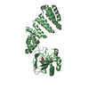
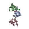
- PDB-5jza: Aspartyl/Asparaginyl beta-hydroxylase (AspH)oxygenase and TPR dom... -
+ Open data

- Basic information
| Entry | Database: PDB / ID: 5jza | ||||||
|---|---|---|---|---|---|---|---|
| Title | Aspartyl/Asparaginyl beta-hydroxylase (AspH)oxygenase and TPR domains in complex with manganese and N-oxalylglycine | ||||||
 Components Components | Aspartyl/asparaginyl beta-hydroxylase | ||||||
 Keywords Keywords |  OXIDOREDUCTASE / 2-oxoglutarate dependent oxygenase / aspartyl/asparaginyl beta-hydroxylase / EGF-like domain hydroxylase / double stranded beta-helix / tetratricopeptide repeat OXIDOREDUCTASE / 2-oxoglutarate dependent oxygenase / aspartyl/asparaginyl beta-hydroxylase / EGF-like domain hydroxylase / double stranded beta-helix / tetratricopeptide repeat | ||||||
| Function / homology |  Function and homology informationpeptide-aspartate beta-dioxygenase / regulation of inositol 1,4,5-trisphosphate-sensitive calcium-release channel activity / regulation of protein depolymerization / activation of store-operated calcium channel activity / regulation of cell communication by electrical coupling / junctional sarcoplasmic reticulum membrane / peptidyl-aspartic acid 3-dioxygenase activity / sarcoplasmic reticulum lumen / limb morphogenesis / cortical endoplasmic reticulum ...peptide-aspartate beta-dioxygenase / regulation of inositol 1,4,5-trisphosphate-sensitive calcium-release channel activity / regulation of protein depolymerization / activation of store-operated calcium channel activity / regulation of cell communication by electrical coupling / junctional sarcoplasmic reticulum membrane / peptidyl-aspartic acid 3-dioxygenase activity / sarcoplasmic reticulum lumen / limb morphogenesis / cortical endoplasmic reticulum / pattern specification process / positive regulation of intracellular protein transport / activation of cysteine-type endopeptidase activity / face morphogenesis / structural constituent of muscle / positive regulation of calcium ion transport into cytosol / response to ATP / roof of mouth development / positive regulation of ryanodine-sensitive calcium-release channel activity / Protein hydroxylation / positive regulation of proteolysis / detection of calcium ion / calcium ion homeostasis / regulation of cardiac muscle contraction by regulation of the release of sequestered calcium ion / regulation of release of sequestered calcium ion into cytosol by sarcoplasmic reticulum / Ion homeostasis / regulation of ryanodine-sensitive calcium-release channel activity / sarcoplasmic reticulum membrane / calcium channel complex / cellular response to calcium ion / muscle contraction / calcium ion transmembrane transport / regulation of protein stability / Stimuli-sensing channels / cell population proliferation / transmembrane transporter binding / electron transfer activity / negative regulation of cell population proliferation / calcium ion binding / endoplasmic reticulum membrane / structural molecule activity / positive regulation of DNA-templated transcription / endoplasmic reticulum / plasma membrane Function and homology informationpeptide-aspartate beta-dioxygenase / regulation of inositol 1,4,5-trisphosphate-sensitive calcium-release channel activity / regulation of protein depolymerization / activation of store-operated calcium channel activity / regulation of cell communication by electrical coupling / junctional sarcoplasmic reticulum membrane / peptidyl-aspartic acid 3-dioxygenase activity / sarcoplasmic reticulum lumen / limb morphogenesis / cortical endoplasmic reticulum ...peptide-aspartate beta-dioxygenase / regulation of inositol 1,4,5-trisphosphate-sensitive calcium-release channel activity / regulation of protein depolymerization / activation of store-operated calcium channel activity / regulation of cell communication by electrical coupling / junctional sarcoplasmic reticulum membrane / peptidyl-aspartic acid 3-dioxygenase activity / sarcoplasmic reticulum lumen / limb morphogenesis / cortical endoplasmic reticulum / pattern specification process / positive regulation of intracellular protein transport / activation of cysteine-type endopeptidase activity / face morphogenesis / structural constituent of muscle / positive regulation of calcium ion transport into cytosol / response to ATP / roof of mouth development / positive regulation of ryanodine-sensitive calcium-release channel activity / Protein hydroxylation / positive regulation of proteolysis / detection of calcium ion / calcium ion homeostasis / regulation of cardiac muscle contraction by regulation of the release of sequestered calcium ion / regulation of release of sequestered calcium ion into cytosol by sarcoplasmic reticulum / Ion homeostasis / regulation of ryanodine-sensitive calcium-release channel activity / sarcoplasmic reticulum membrane / calcium channel complex / cellular response to calcium ion / muscle contraction / calcium ion transmembrane transport / regulation of protein stability / Stimuli-sensing channels / cell population proliferation / transmembrane transporter binding / electron transfer activity / negative regulation of cell population proliferation / calcium ion binding / endoplasmic reticulum membrane / structural molecule activity / positive regulation of DNA-templated transcription / endoplasmic reticulum / plasma membraneSimilarity search - Function | ||||||
| Biological species |  Homo sapiens (human) Homo sapiens (human) | ||||||
| Method | X-RAY DIFFRACTION / SYNCHROTRON / MOLECULAR REPLACEMENT / Resolution: 2.14 Å | ||||||
 Authors Authors | McDonough, M.A. / Pfeffer, I. | ||||||
 Citation Citation | Journal: Nat Commun / Year: 2019 Title: Aspartate/asparagine-beta-hydroxylase crystal structures reveal an unexpected epidermal growth factor-like domain substrate disulfide pattern. Authors: Pfeffer, I. / Brewitz, L. / Krojer, T. / Jensen, S.A. / Kochan, G.T. / Kershaw, N.J. / Hewitson, K.S. / McNeill, L.A. / Kramer, H. / Munzel, M. / Hopkinson, R.J. / Oppermann, U. / Handford, ...Authors: Pfeffer, I. / Brewitz, L. / Krojer, T. / Jensen, S.A. / Kochan, G.T. / Kershaw, N.J. / Hewitson, K.S. / McNeill, L.A. / Kramer, H. / Munzel, M. / Hopkinson, R.J. / Oppermann, U. / Handford, P.A. / McDonough, M.A. / Schofield, C.J. | ||||||
| History |
|
- Structure visualization
| Structure viewer | Molecule: MolmilJmol/JSmol |
|---|
- Downloads & links
-Download
| PDBx/mmCIF format | 5jza.cif.gz | 195.7 KB | Display | PDBx/mmCIF format |
|---|---|---|---|---|
| PDB format | pdb5jza.ent.gz | 153.9 KB | Display | PDB format |
| PDBx/mmJSON format | 5jza.json.gz | Tree view | PDBx/mmJSON format | |
| Others |  Other downloads Other downloads |
-Validation report
| Arichive directory | https://data.pdbj.org/pub/pdb/validation_reports/jz/5jzaftp://data.pdbj.org/pub/pdb/validation_reports/jz/5jza | HTTPS FTP |
|---|
-Related structure data
| Related structure data | 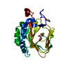 5apaSC  5jqyC 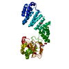 5jz6C 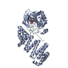 5jz8C 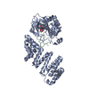 5jzuC 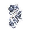 6rk9C S: Starting model for refinement C: citing same article ( |
|---|---|
| Similar structure data |


-Links
 PDBj
PDBj


- Assembly
| Deposited unit | 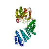
| ||||||||
|---|---|---|---|---|---|---|---|---|---|
| 1 |
| ||||||||
| Unit cell |
|
-Components
| #1: Protein | Mass: 49405.336 Da / Num. of mol.: 1 Source method: isolated from a genetically manipulated source Source: (gene. exp.) Homo sapiens (human) / Gene: ASPH, BAH / Plasmid: pET-28a(+) / Production host:  Escherichia coli BL21(DE3) (bacteria) Escherichia coli BL21(DE3) (bacteria)References: UniProt: Q12797, peptide-aspartate beta-dioxygenase | ||||
|---|---|---|---|---|---|
| #2: Chemical | ChemComp-MN /   Mass: 54.938 Da / Num. of mol.: 1 / Source method: obtained synthetically / Formula: Mn Mass: 54.938 Da / Num. of mol.: 1 / Source method: obtained synthetically / Formula: Mn | ||||
| #3: Chemical | ChemComp-GOL / Glycerol  Mass: 92.094 Da / Num. of mol.: 4 / Source method: obtained synthetically / Formula: C3H8O3 Mass: 92.094 Da / Num. of mol.: 4 / Source method: obtained synthetically / Formula: C3H8O3#4: Chemical | ChemComp-OGA / | N-Oxalylglycine  Mass: 147.086 Da / Num. of mol.: 1 / Source method: obtained synthetically / Formula: C4H5NO5 / Comment: inhibitor*YM Mass: 147.086 Da / Num. of mol.: 1 / Source method: obtained synthetically / Formula: C4H5NO5 / Comment: inhibitor*YM#5: Water | ChemComp-HOH / | Water Mass: 18.015 Da / Num. of mol.: 125 / Source method: isolated from a natural source / Formula: H2O Mass: 18.015 Da / Num. of mol.: 125 / Source method: isolated from a natural source / Formula: H2O |
-Experimental details
-Experiment
| Experiment | Method: X-RAY DIFFRACTION / Number of used crystals: 1 |
|---|
- Sample preparation
| Crystal | Density Matthews: 2.95 Å3/Da / Density % sol: 58.33 % |
|---|---|
| Crystal grow | Temperature: 277 K / Method: vapor diffusion, sitting drop / pH: 9 Details: 100 mM bicine, 100 mM sodium chloride, 27% PEG 550 MME, 1 mM manganese chloride, 2 mM N-oxalylglycine, 18 mg/ml protein |
-Data collection
| Diffraction | Mean temperature: 100 K |
|---|---|
| Diffraction source | Source: SYNCHROTRON / Site: Diamond  / Beamline: I02 / Wavelength: 0.9795 Å / Beamline: I02 / Wavelength: 0.9795 Å |
| Detector | Type: DECTRIS PILATUS 6M-F / Detector: PIXEL / Date: Jul 5, 2012 |
| Radiation | Protocol: SINGLE WAVELENGTH / Monochromatic (M) / Laue (L): M / Scattering type: x-ray |
| Radiation wavelength | Wavelength: 0.9795 Å / Relative weight: 1 |
| Reflection | Resolution: 2.14→85.21 Å / Num. obs: 33179 / % possible obs: 99.9 % / Redundancy: 6.4 % / Biso Wilson estimate: 46.25 Å2 / Rmerge(I) obs: 0.049 / Net I/σ(I): 18.6 |
| Reflection shell | Resolution: 2.14→2.2 Å / Redundancy: 6.8 % / Rmerge(I) obs: 0.652 / Mean I/σ(I) obs: 3.1 / % possible all: 99.9 |
- Processing
| Software |
| ||||||||||||||||||||||||||||||||||||||||||||||||||||||||||||||||||||||||||||||||||||||||||||||||||||||||||||||||||||||||||||||||||||||||||||||||||||||||||||||||||||||||||||||||||||||||||||||||||||
|---|---|---|---|---|---|---|---|---|---|---|---|---|---|---|---|---|---|---|---|---|---|---|---|---|---|---|---|---|---|---|---|---|---|---|---|---|---|---|---|---|---|---|---|---|---|---|---|---|---|---|---|---|---|---|---|---|---|---|---|---|---|---|---|---|---|---|---|---|---|---|---|---|---|---|---|---|---|---|---|---|---|---|---|---|---|---|---|---|---|---|---|---|---|---|---|---|---|---|---|---|---|---|---|---|---|---|---|---|---|---|---|---|---|---|---|---|---|---|---|---|---|---|---|---|---|---|---|---|---|---|---|---|---|---|---|---|---|---|---|---|---|---|---|---|---|---|---|---|---|---|---|---|---|---|---|---|---|---|---|---|---|---|---|---|---|---|---|---|---|---|---|---|---|---|---|---|---|---|---|---|---|---|---|---|---|---|---|---|---|---|---|---|---|---|---|---|---|
| Refinement | Method to determine structure: MOLECULAR REPLACEMENT Starting model: 5APA Resolution: 2.14→85.21 Å / SU ML: 0.23 / Cross valid method: FREE R-VALUE / σ(F): 1.34 / Phase error: 20.07
| ||||||||||||||||||||||||||||||||||||||||||||||||||||||||||||||||||||||||||||||||||||||||||||||||||||||||||||||||||||||||||||||||||||||||||||||||||||||||||||||||||||||||||||||||||||||||||||||||||||
| Solvent computation | Shrinkage radii: 0.9 Å / VDW probe radii: 1.11 Å | ||||||||||||||||||||||||||||||||||||||||||||||||||||||||||||||||||||||||||||||||||||||||||||||||||||||||||||||||||||||||||||||||||||||||||||||||||||||||||||||||||||||||||||||||||||||||||||||||||||
| Displacement parameters | Biso max: 183.18 Å2 / Biso mean: 68.3722 Å2 / Biso min: 31.28 Å2 | ||||||||||||||||||||||||||||||||||||||||||||||||||||||||||||||||||||||||||||||||||||||||||||||||||||||||||||||||||||||||||||||||||||||||||||||||||||||||||||||||||||||||||||||||||||||||||||||||||||
| Refinement step | Cycle: final / Resolution: 2.14→85.21 Å
| ||||||||||||||||||||||||||||||||||||||||||||||||||||||||||||||||||||||||||||||||||||||||||||||||||||||||||||||||||||||||||||||||||||||||||||||||||||||||||||||||||||||||||||||||||||||||||||||||||||
| Refine LS restraints |
| ||||||||||||||||||||||||||||||||||||||||||||||||||||||||||||||||||||||||||||||||||||||||||||||||||||||||||||||||||||||||||||||||||||||||||||||||||||||||||||||||||||||||||||||||||||||||||||||||||||
| LS refinement shell | Refine-ID: X-RAY DIFFRACTION / Total num. of bins used: 27
| ||||||||||||||||||||||||||||||||||||||||||||||||||||||||||||||||||||||||||||||||||||||||||||||||||||||||||||||||||||||||||||||||||||||||||||||||||||||||||||||||||||||||||||||||||||||||||||||||||||
| Refinement TLS params. | Method: refined / Refine-ID: X-RAY DIFFRACTION
| ||||||||||||||||||||||||||||||||||||||||||||||||||||||||||||||||||||||||||||||||||||||||||||||||||||||||||||||||||||||||||||||||||||||||||||||||||||||||||||||||||||||||||||||||||||||||||||||||||||
| Refinement TLS group |
|
