 Movie
Movie Controller


+ Open data

- Basic information
| Entry | Database: PDB / ID: 5juy | ||||||
|---|---|---|---|---|---|---|---|
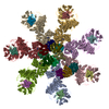
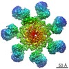
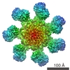
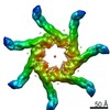
| Title | Active human apoptosome with procaspase-9 | ||||||
 Components Components |
| ||||||
 Keywords Keywords |  APOPTOSIS / Apaf-1 / programmed cell death / AAA+ ATPase APOPTOSIS / Apaf-1 / programmed cell death / AAA+ ATPase | ||||||
| Function / homology |  Function and homology information Function and homology informationRelease of apoptotic factors from the mitochondria / Formation of apoptosome / Activation of caspases through apoptosome-mediated cleavage / Pyroptosis / Regulation of the apoptosome activity / caspase-9 / Transcriptional activation of mitochondrial biogenesis / response to G1 DNA damage checkpoint signaling / caspase complex / Detoxification of Reactive Oxygen Species ...Release of apoptotic factors from the mitochondria / Formation of apoptosome / Activation of caspases through apoptosome-mediated cleavage / Pyroptosis / Regulation of the apoptosome activity / caspase-9 / Transcriptional activation of mitochondrial biogenesis / response to G1 DNA damage checkpoint signaling / caspase complex / Detoxification of Reactive Oxygen Species / regulation of apoptotic DNA fragmentation / Formation of apoptosome / apoptosome / activation of cysteine-type endopeptidase activity involved in apoptotic process by cytochrome c / TP53 Regulates Metabolic Genes / Cytoprotection by HMOX1 / leukocyte apoptotic process / glial cell apoptotic process / Respiratory electron transport / response to cobalt ion / cysteine-type endopeptidase activity involved in apoptotic signaling pathway / Caspase activation via Dependence Receptors in the absence of ligand / Regulation of the apoptosome activity / Activation of caspases through apoptosome-mediated cleavage / cysteine-type endopeptidase activity involved in apoptotic process / SMAC (DIABLO) binds to IAPs / SMAC(DIABLO)-mediated dissociation of IAP:caspase complexes / mitochondrial electron transport, cytochrome c to oxygen / fibroblast apoptotic process / AKT phosphorylates targets in the cytosol / epithelial cell apoptotic process / mitochondrial electron transport, ubiquinol to cytochrome c / platelet formation / TP53 Regulates Transcription of Caspase Activators and Caspases / Transcriptional Regulation by E2F6 / Constitutive Signaling by AKT1 E17K in Cancer / cysteine-type endopeptidase activator activity involved in apoptotic process / protein maturation / enzyme activator activity / intrinsic apoptotic signaling pathway in response to endoplasmic reticulum stress / respirasome / signal transduction in response to DNA damage / forebrain development / positive regulation of apoptotic signaling pathway / cardiac muscle cell apoptotic process / cellular response to transforming growth factor beta stimulus / heat shock protein binding / cellular response to dexamethasone stimulus / intrinsic apoptotic signaling pathway / response to nutrient / kidney development / neural tube closure / response to ischemia / ADP binding / NOD1/2 Signaling Pathway / mitochondrial intermembrane space / protein processing / SH3 domain binding / activation of cysteine-type endopeptidase activity involved in apoptotic process / positive regulation of neuron apoptotic process / cellular response to UV / intrinsic apoptotic signaling pathway in response to DNA damage / response to estradiol / nervous system development / peptidase activity / neuron apoptotic process / regulation of apoptotic process / secretory granule lumen / ficolin-1-rich granule lumen / response to lipopolysaccharide / cell differentiation / electron transfer activity / response to hypoxia / positive regulation of apoptotic process / cysteine-type endopeptidase activity / nucleotide binding / apoptotic process / DNA damage response / heme binding / Neutrophil degranulation / protein kinase binding / protein-containing complex / mitochondrion / extracellular exosome / extracellular region / ATP binding / identical protein binding / metal ion binding / nucleus / cytosol / cytoplasmSimilarity search - Function | ||||||
| Biological species |  Homo sapiens (human) Homo sapiens (human) Bos taurus (cattle) Bos taurus (cattle) | ||||||
| Method | ELECTRON MICROSCOPY / single particle reconstruction / cryo EM / Resolution: 4.1 Å | ||||||
 Authors Authors | Cheng, T.C. / Hong, C. / Akey, I.V. / Yuan, S. / Akey, C.W. | ||||||
| Funding support |  United States, 1items United States, 1items
| ||||||
 Citation Citation | Journal: Elife / Year: 2016 Title: A near atomic structure of the active human apoptosome. Authors: Tat Cheung Cheng / Chuan Hong / Ildikó V Akey / Shujun Yuan / Christopher W Akey / Abstract: In response to cell death signals, an active apoptosome is assembled from Apaf-1 and procaspase-9 (pc-9). Here we report a near atomic structure of the active human apoptosome determined by cryo- ...In response to cell death signals, an active apoptosome is assembled from Apaf-1 and procaspase-9 (pc-9). Here we report a near atomic structure of the active human apoptosome determined by cryo-electron microscopy. The resulting model gives insights into cytochrome c binding, nucleotide exchange and conformational changes that drive assembly. During activation an acentric disk is formed on the central hub of the apoptosome. This disk contains four Apaf-1/pc-9 CARD pairs arranged in a shallow spiral with the fourth pc-9 CARD at lower occupancy. On average, Apaf-1 CARDs recruit 3 to 5 pc-9 molecules to the apoptosome and one catalytic domain may be parked on the hub, when an odd number of zymogens are bound. This suggests a stoichiometry of one or at most, two pc-9 dimers per active apoptosome. Thus, our structure provides a molecular framework to understand the role of the apoptosome in programmed cell death and disease. | ||||||
| History |
|
- Structure visualization
| Movie |
Movie viewer |
|---|---|
| Structure viewer | Molecule: MolmilJmol/JSmol |
- Downloads & links
-Download
| PDBx/mmCIF format | 5juy.cif.gz | 1.6 MB | Display | PDBx/mmCIF format |
|---|---|---|---|---|
| PDB format | pdb5juy.ent.gz | 1.3 MB | Display | PDB format |
| PDBx/mmJSON format | 5juy.json.gz | Tree view | PDBx/mmJSON format | |
| Others |  Other downloads Other downloads |
-Validation report
| Arichive directory | https://data.pdbj.org/pub/pdb/validation_reports/ju/5juyftp://data.pdbj.org/pub/pdb/validation_reports/ju/5juy | HTTPS FTP |
|---|
-Related structure data
| Related structure data | 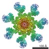 8178MC M: map data used to model this data C: citing same article ( |
|---|---|
| Similar structure data |

-Links
 PDBj
PDBj


- Assembly
| Deposited unit | 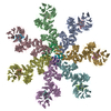
|
|---|---|
| 1 |
|
-Components
| #1: Protein | Mass: 142023.672 Da / Num. of mol.: 7 Source method: isolated from a genetically manipulated source Details: without N-terminal CARDs / Source: (gene. exp.) Homo sapiens (human) / Gene: APAF1, KIAA0413 / Production host:   Spodoptera frugiperda (fall armyworm) / References: UniProt: O14727 Spodoptera frugiperda (fall armyworm) / References: UniProt: O14727#2: Protein | Mass: 11595.392 Da / Num. of mol.: 7 / Source method: isolated from a natural source / Source: (natural) Bos taurus (cattle) / References: UniProt: P62894#3: Protein | / CASP-9 / Apoptotic protease Mch-6 / Apoptotic protease-activating factor 3 / APAF-3 / ICE-like ...CASP-9 / Apoptotic protease Mch-6 / Apoptotic protease-activating factor 3 / APAF-3 / ICE-like apoptotic protease 6 / ICE-LAP6Mass: 11140.723 Da / Num. of mol.: 4 / Fragment: N-terminal caspase recognition domain Source method: isolated from a genetically manipulated source Details: N-terminal caspase recognition domain (CARD) of procaspase-9 Source: (gene. exp.) Homo sapiens (human) / Gene: CASP9, MCH6Production host:  Escherichia coli 'BL21-Gold(DE3)pLysS AG' (bacteria) Escherichia coli 'BL21-Gold(DE3)pLysS AG' (bacteria)References: UniProt: P55211, caspase-9#4: Chemical | ChemComp-DTP / Deoxyadenosine triphosphate  Mass: 491.182 Da / Num. of mol.: 7 / Source method: obtained synthetically / Formula: C10H16N5O12P3 Mass: 491.182 Da / Num. of mol.: 7 / Source method: obtained synthetically / Formula: C10H16N5O12P3#5: Chemical | ChemComp-HEC / Heme C  Mass: 618.503 Da / Num. of mol.: 7 / Source method: obtained synthetically / Formula: C34H34FeN4O4 Mass: 618.503 Da / Num. of mol.: 7 / Source method: obtained synthetically / Formula: C34H34FeN4O4 |
|---|
-Experimental details
-Experiment
| Experiment | Method: ELECTRON MICROSCOPY |
|---|---|
| EM experiment | Aggregation state: PARTICLE / 3D reconstruction method: single particle reconstruction |
- Sample preparation
| Component | Name: Active complex of Apaf-1, cytochrome c and pro-caspase 9 Type: COMPLEX Details: Complex stoichiometry: 4 full length Apaf-1 molecules with ordered N-terminal CARDs, 3 truncated Apaf-1 molecules without CARDs, 7 cytochrome c molecules and 4 N-terminal CARDs from procaspase-9 Entity ID: #1-#4 / Source: RECOMBINANT | |||||||||||||||||||||||||||||||||||
|---|---|---|---|---|---|---|---|---|---|---|---|---|---|---|---|---|---|---|---|---|---|---|---|---|---|---|---|---|---|---|---|---|---|---|---|---|
| Molecular weight | Value: 1.3 MDa / Experimental value: NO | |||||||||||||||||||||||||||||||||||
| Source (natural) | Organism: Homo sapiens (human) | |||||||||||||||||||||||||||||||||||
| Source (recombinant) | Organism: Spodoptera frugiperda (fall armyworm) / Plasmid: pFast-Bac | |||||||||||||||||||||||||||||||||||
| Buffer solution | pH: 7.5 / Details: Buffer A | |||||||||||||||||||||||||||||||||||
| Buffer component |
| |||||||||||||||||||||||||||||||||||
| Specimen | Conc.: 4 mg/ml / Embedding applied: NO / Shadowing applied: NO / Staining applied: NO / Vitrification applied: YES | |||||||||||||||||||||||||||||||||||
| Specimen support | Grid material: COPPER / Grid mesh size: 300 divisions/in. / Grid type: C-flat-1.2/1.3 | |||||||||||||||||||||||||||||||||||
| Vitrification | Instrument: FEI VITROBOT MARK III / Cryogen name: ETHANE / Humidity: 100 % / Chamber temperature: 293 K |
- Electron microscopy imaging
| Experimental equipment |  Model: Titan Krios / Image courtesy: FEI Company |
|---|---|
| Microscopy | Model: FEI TITAN KRIOS Details: data collected with a Cs aberration corrector and a GIF energy filter with a slit width of 20 eV. |
| Electron gun | Electron source: FIELD EMISSION GUN / Accelerating voltage: 300 kV / Illumination mode: FLOOD BEAM |
| Electron lens | Mode: BRIGHT FIELDBright-field microscopy / Nominal magnification: 81000 X / Nominal defocus max: 2400 nm / Nominal defocus min: 1500 nm / Cs: 0.01 mm / C2 aperture diameter: 100 µm / Alignment procedure: COMA FREE |
| Specimen holder | Cryogen: NITROGEN / Specimen holder model: FEI TITAN KRIOS AUTOGRID HOLDER |
| Image recording | Average exposure time: 0.3 sec. / Electron dose: 1.8 e/Å2 / Detector mode: SUPER-RESOLUTION / Film or detector model: GATAN K2 SUMMIT (4k x 4k) / Num. of grids imaged: 1 / Num. of real images: 1991 |
| EM imaging optics | Energyfilter name: GIF |
| Image scans | Width: 7676 / Height: 7420 / Movie frames/image: 23 / Used frames/image: 2-23 |
- Processing
| EM software |
| ||||||||||||||||||||||||||||||||||||||||
|---|---|---|---|---|---|---|---|---|---|---|---|---|---|---|---|---|---|---|---|---|---|---|---|---|---|---|---|---|---|---|---|---|---|---|---|---|---|---|---|---|---|
| CTF correction | Type: PHASE FLIPPING AND AMPLITUDE CORRECTION | ||||||||||||||||||||||||||||||||||||||||
| Particle selection | Num. of particles selected: 134970 | ||||||||||||||||||||||||||||||||||||||||
| Symmetry | Point symmetry: C7 (7 fold cyclic) | ||||||||||||||||||||||||||||||||||||||||
| 3D reconstruction | Resolution: 4.1 Å / Resolution method: FSC 0.143 CUT-OFF / Num. of particles: 92867 / Algorithm: FOURIER SPACE / Symmetry type: POINT | ||||||||||||||||||||||||||||||||||||||||
| Atomic model building | Protocol: FLEXIBLE FIT / Space: REAL Details: Initial rigid body fitting with Chimera with a previously published starting model (PDB 3J2T), molecular dynamics flexible fitting with MDFF and real space density refinement with Phenix |
