 Movie
Movie Controller

[English] 日本語
 Yorodumi
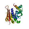
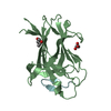
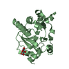
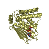
- PDB-5jr2: Crystal structure of the EphA4 LBD in complex with APYd3 peptide ... -
+ Open data

- Basic information
| Entry | Database: PDB / ID: 5jr2 | ||||||||||||
|---|---|---|---|---|---|---|---|---|---|---|---|---|---|
| Title | Crystal structure of the EphA4 LBD in complex with APYd3 peptide inhibitor | ||||||||||||
 Components Components |
| ||||||||||||
 Keywords Keywords | TRANSFERASE/INHIBITOR /  receptor tyrosine kinase / peptide inhibitor / ephrin / ALS / TRANSFERASE-INHIBITOR complex receptor tyrosine kinase / peptide inhibitor / ephrin / ALS / TRANSFERASE-INHIBITOR complex | ||||||||||||
| Function / homology |  Function and homology informationDH domain binding / neuron projection fasciculation / positive regulation of Rho guanyl-nucleotide exchange factor activity / corticospinal tract morphogenesis / regulation of astrocyte differentiation / negative regulation of proteolysis involved in protein catabolic process / neuron projection guidance / nephric duct morphogenesis / regulation of synapse pruning / fasciculation of sensory neuron axon ...DH domain binding / neuron projection fasciculation / positive regulation of Rho guanyl-nucleotide exchange factor activity / corticospinal tract morphogenesis / regulation of astrocyte differentiation / negative regulation of proteolysis involved in protein catabolic process / neuron projection guidance / nephric duct morphogenesis / regulation of synapse pruning / fasciculation of sensory neuron axon / fasciculation of motor neuron axon / synapse pruning / negative regulation of cellular response to hypoxia / negative regulation of axon regeneration / positive regulation of aspartic-type endopeptidase activity involved in amyloid precursor protein catabolic process / glial cell migration / transmembrane-ephrin receptor activity / PH domain binding / GPI-linked ephrin receptor activity / regulation of modification of synaptic structure / negative regulation of epithelial to mesenchymal transition / regulation of dendritic spine morphogenesis / negative regulation of cell adhesion / motor neuron axon guidance / adult walking behavior / adherens junction organization / positive regulation of dendrite morphogenesis / EPH-Ephrin signaling / Somitogenesis / regulation of axonogenesis / positive regulation of amyloid-beta formation / cochlea development / regulation of GTPase activity / EPHA-mediated growth cone collapse / negative regulation of long-term synaptic potentiation / positive regulation of cell adhesion / EPH-ephrin mediated repulsion of cells / ephrin receptor signaling pathway / positive regulation of protein tyrosine kinase activity / axon terminus / axonal growth cone / protein tyrosine kinase binding / ephrin receptor binding / negative regulation of cell migration / dendritic shaft / filopodium / axon guidance / postsynaptic density membrane / adherens junction / Schaffer collateral - CA1 synapse / neuromuscular junction / negative regulation of ERK1 and ERK2 cascade / receptor protein-tyrosine kinase / peptidyl-tyrosine phosphorylation / cellular response to amyloid-beta / negative regulation of neuron projection development / presynaptic membrane / amyloid-beta binding / kinase activity / perikaryon / early endosome membrane / protein tyrosine kinase activity / mitochondrial outer membrane / dendritic spine / protein autophosphorylation / negative regulation of translation / protein stabilization / cell adhesion / protein kinase activity / positive regulation of cell migration / axon / glutamatergic synapse / dendrite / positive regulation of cell population proliferation / cell surface / ATP binding / identical protein binding / plasma membrane / cytoplasm Function and homology informationDH domain binding / neuron projection fasciculation / positive regulation of Rho guanyl-nucleotide exchange factor activity / corticospinal tract morphogenesis / regulation of astrocyte differentiation / negative regulation of proteolysis involved in protein catabolic process / neuron projection guidance / nephric duct morphogenesis / regulation of synapse pruning / fasciculation of sensory neuron axon ...DH domain binding / neuron projection fasciculation / positive regulation of Rho guanyl-nucleotide exchange factor activity / corticospinal tract morphogenesis / regulation of astrocyte differentiation / negative regulation of proteolysis involved in protein catabolic process / neuron projection guidance / nephric duct morphogenesis / regulation of synapse pruning / fasciculation of sensory neuron axon / fasciculation of motor neuron axon / synapse pruning / negative regulation of cellular response to hypoxia / negative regulation of axon regeneration / positive regulation of aspartic-type endopeptidase activity involved in amyloid precursor protein catabolic process / glial cell migration / transmembrane-ephrin receptor activity / PH domain binding / GPI-linked ephrin receptor activity / regulation of modification of synaptic structure / negative regulation of epithelial to mesenchymal transition / regulation of dendritic spine morphogenesis / negative regulation of cell adhesion / motor neuron axon guidance / adult walking behavior / adherens junction organization / positive regulation of dendrite morphogenesis / EPH-Ephrin signaling / Somitogenesis / regulation of axonogenesis / positive regulation of amyloid-beta formation / cochlea development / regulation of GTPase activity / EPHA-mediated growth cone collapse / negative regulation of long-term synaptic potentiation / positive regulation of cell adhesion / EPH-ephrin mediated repulsion of cells / ephrin receptor signaling pathway / positive regulation of protein tyrosine kinase activity / axon terminus / axonal growth cone / protein tyrosine kinase binding / ephrin receptor binding / negative regulation of cell migration / dendritic shaft / filopodium / axon guidance / postsynaptic density membrane / adherens junction / Schaffer collateral - CA1 synapse / neuromuscular junction / negative regulation of ERK1 and ERK2 cascade / receptor protein-tyrosine kinase / peptidyl-tyrosine phosphorylation / cellular response to amyloid-beta / negative regulation of neuron projection development / presynaptic membrane / amyloid-beta binding / kinase activity / perikaryon / early endosome membrane / protein tyrosine kinase activity / mitochondrial outer membrane / dendritic spine / protein autophosphorylation / negative regulation of translation / protein stabilization / cell adhesion / protein kinase activity / positive regulation of cell migration / axon / glutamatergic synapse / dendrite / positive regulation of cell population proliferation / cell surface / ATP binding / identical protein binding / plasma membrane / cytoplasmSimilarity search - Function | ||||||||||||
| Biological species |  Homo sapiens (human) Homo sapiens (human)synthetic construct (others) | ||||||||||||
| Method | X-RAY DIFFRACTION / MOLECULAR REPLACEMENT / Resolution: 1.75 Å | ||||||||||||
 Authors Authors | Lechtenberg, B.C. / Olson, E.J. / Pasquale, E.B. / Dawson, P.E. / Riedl, S.J. | ||||||||||||
| Funding support |  United States, 3items United States, 3items
| ||||||||||||
 Citation Citation | Journal: Acs Med.Chem.Lett. / Year: 2016 Title: Modifications of a Nanomolar Cyclic Peptide Antagonist for the EphA4 Receptor To Achieve High Plasma Stability. Authors: Olson, E.J. / Lechtenberg, B.C. / Zhao, C. / Rubio de la Torre, E. / Lamberto, I. / Riedl, S.J. / Dawson, P.E. / Pasquale, E.B. | ||||||||||||
| History |
|
- Structure visualization
| Structure viewer | Molecule: MolmilJmol/JSmol |
|---|
- Downloads & links
-Download
| PDBx/mmCIF format | 5jr2.cif.gz | 344.8 KB | Display | PDBx/mmCIF format |
|---|---|---|---|---|
| PDB format | pdb5jr2.ent.gz | 282.6 KB | Display | PDB format |
| PDBx/mmJSON format | 5jr2.json.gz | Tree view | PDBx/mmJSON format | |
| Others |  Other downloads Other downloads |
-Validation report
| Arichive directory | https://data.pdbj.org/pub/pdb/validation_reports/jr/5jr2ftp://data.pdbj.org/pub/pdb/validation_reports/jr/5jr2 | HTTPS FTP |
|---|
-Related structure data
| Related structure data |  4w4zS S: Starting model for refinement |
|---|---|
| Similar structure data |


-Links
 PDBj
PDBj


- Assembly
| Deposited unit | 
| ||||||||||||||||||||||||||||||||||||||||||||||||||||||||||||||||||||||||||||||||||||||||||||||||||
|---|---|---|---|---|---|---|---|---|---|---|---|---|---|---|---|---|---|---|---|---|---|---|---|---|---|---|---|---|---|---|---|---|---|---|---|---|---|---|---|---|---|---|---|---|---|---|---|---|---|---|---|---|---|---|---|---|---|---|---|---|---|---|---|---|---|---|---|---|---|---|---|---|---|---|---|---|---|---|---|---|---|---|---|---|---|---|---|---|---|---|---|---|---|---|---|---|---|---|---|
| 1 | 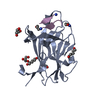
| ||||||||||||||||||||||||||||||||||||||||||||||||||||||||||||||||||||||||||||||||||||||||||||||||||
| 2 | 
| ||||||||||||||||||||||||||||||||||||||||||||||||||||||||||||||||||||||||||||||||||||||||||||||||||
| 3 | 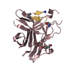
| ||||||||||||||||||||||||||||||||||||||||||||||||||||||||||||||||||||||||||||||||||||||||||||||||||
| 4 | 
| ||||||||||||||||||||||||||||||||||||||||||||||||||||||||||||||||||||||||||||||||||||||||||||||||||
| Unit cell |
| ||||||||||||||||||||||||||||||||||||||||||||||||||||||||||||||||||||||||||||||||||||||||||||||||||
| Noncrystallographic symmetry (NCS) | NCS domain:
NCS domain segments: Component-ID: 0 / Beg auth comp-ID: GLY / Beg label comp-ID: GLY / End auth comp-ID: ALA / End label comp-ID: ALA / Refine code: 0 / Auth seq-ID: 26 - 204 / Label seq-ID: 1 - 179
NCS ensembles :
|
-Components
| #1: Protein | Mass: 20489.143 Da / Num. of mol.: 4 / Mutation: C204A Source method: isolated from a genetically manipulated source Source: (gene. exp.) Homo sapiens (human) / Gene: EPHA4, HEK8, SEK, TYRO1 / Production host:  Escherichia coli (E. coli) Escherichia coli (E. coli)References: UniProt: P54764, receptor protein-tyrosine kinase#2: Protein/peptide | Mass: 1404.617 Da / Num. of mol.: 4 / Source method: obtained synthetically / Source: (synth.) synthetic construct (others) #3: Chemical | ChemComp-HEZ / 1,6-Hexanediol  Mass: 118.174 Da / Num. of mol.: 6 / Source method: obtained synthetically / Formula: C6H14O2 Mass: 118.174 Da / Num. of mol.: 6 / Source method: obtained synthetically / Formula: C6H14O2#4: Chemical | ChemComp-GOL / Glycerol  Mass: 92.094 Da / Num. of mol.: 8 / Source method: obtained synthetically / Formula: C3H8O3 Mass: 92.094 Da / Num. of mol.: 8 / Source method: obtained synthetically / Formula: C3H8O3#5: Water | ChemComp-HOH / | Water Mass: 18.015 Da / Num. of mol.: 685 / Source method: isolated from a natural source / Formula: H2O Mass: 18.015 Da / Num. of mol.: 685 / Source method: isolated from a natural source / Formula: H2O |
|---|
-Experimental details
-Experiment
| Experiment | Method: X-RAY DIFFRACTION / Number of used crystals: 1 |
|---|
- Sample preparation
| Crystal | Density Matthews: 2.29 Å3/Da / Density % sol: 46.3 % |
|---|---|
| Crystal grow | Temperature: 293 K / Method: vapor diffusion, sitting drop / pH: 6.5 Details: 0.2 M MgCl2, 0.1 M Bis-Tris pH 6.5, 25% PEG 3,350, 3% 1,6-hexanediol |
-Data collection
| Diffraction | Mean temperature: 100 K |
|---|---|
| Diffraction source | Source: ROTATING ANODE / Type: RIGAKU FR-E SUPERBRIGHT / Wavelength: 1.5418 Å |
| Detector | Type: RIGAKU RAXIS HTC / Detector: IMAGE PLATE / Date: Dec 4, 2015 |
| Radiation | Protocol: SINGLE WAVELENGTH / Monochromatic (M) / Laue (L): M / Scattering type: x-ray |
| Radiation wavelength | Wavelength: 1.5418 Å / Relative weight: 1 |
| Reflection | Resolution: 1.75→28.92 Å / Num. obs: 78825 / % possible obs: 99.3 % / Redundancy: 3.6 % / Biso Wilson estimate: 17.1 Å2 / CC1/2: 0.999 / Rmerge(I) obs: 0.042 / Net I/σ(I): 17.9 |
| Reflection shell | Resolution: 1.75→1.78 Å / Redundancy: 3.1 % / Rmerge(I) obs: 0.316 / Mean I/σ(I) obs: 3.1 / % possible all: 96.5 |
- Processing
| Software |
| ||||||||||||||||||||||||||||||||||||||||||||||||||||||||||||||||||||||||||||||||||||||||||||||||||||||||||||||||||||||||||||||||||||||||||||||||||||||||||||||||||||||||||||||||||||||
|---|---|---|---|---|---|---|---|---|---|---|---|---|---|---|---|---|---|---|---|---|---|---|---|---|---|---|---|---|---|---|---|---|---|---|---|---|---|---|---|---|---|---|---|---|---|---|---|---|---|---|---|---|---|---|---|---|---|---|---|---|---|---|---|---|---|---|---|---|---|---|---|---|---|---|---|---|---|---|---|---|---|---|---|---|---|---|---|---|---|---|---|---|---|---|---|---|---|---|---|---|---|---|---|---|---|---|---|---|---|---|---|---|---|---|---|---|---|---|---|---|---|---|---|---|---|---|---|---|---|---|---|---|---|---|---|---|---|---|---|---|---|---|---|---|---|---|---|---|---|---|---|---|---|---|---|---|---|---|---|---|---|---|---|---|---|---|---|---|---|---|---|---|---|---|---|---|---|---|---|---|---|---|---|
| Refinement | Method to determine structure: MOLECULAR REPLACEMENT Starting model: 4W4Z Resolution: 1.75→28.92 Å / Cor.coef. Fo:Fc: 0.958 / Cor.coef. Fo:Fc free: 0.942 / SU B: 4.697 / SU ML: 0.078 / Cross valid method: THROUGHOUT / ESU R: 0.121 / ESU R Free: 0.113 / Details: HYDROGENS HAVE BEEN ADDED IN THE RIDING POSITIONS
| ||||||||||||||||||||||||||||||||||||||||||||||||||||||||||||||||||||||||||||||||||||||||||||||||||||||||||||||||||||||||||||||||||||||||||||||||||||||||||||||||||||||||||||||||||||||
| Solvent computation | Ion probe radii: 0.8 Å / Shrinkage radii: 0.8 Å / VDW probe radii: 1 Å | ||||||||||||||||||||||||||||||||||||||||||||||||||||||||||||||||||||||||||||||||||||||||||||||||||||||||||||||||||||||||||||||||||||||||||||||||||||||||||||||||||||||||||||||||||||||
| Displacement parameters | Biso mean: 22.711 Å2
| ||||||||||||||||||||||||||||||||||||||||||||||||||||||||||||||||||||||||||||||||||||||||||||||||||||||||||||||||||||||||||||||||||||||||||||||||||||||||||||||||||||||||||||||||||||||
| Refinement step | Cycle: 1 / Resolution: 1.75→28.92 Å
| ||||||||||||||||||||||||||||||||||||||||||||||||||||||||||||||||||||||||||||||||||||||||||||||||||||||||||||||||||||||||||||||||||||||||||||||||||||||||||||||||||||||||||||||||||||||
| Refine LS restraints |
|