 Movie
Movie Controller


+ Open data

- Basic information
| Entry | Database: PDB / ID: 5jou | ||||||||||||
|---|---|---|---|---|---|---|---|---|---|---|---|---|---|
| Title | Bacteroides ovatus Xyloglucan PUL GH31 | ||||||||||||
 Components Components | Alpha-xylosidase BoGH31A | ||||||||||||
 Keywords Keywords |  HYDROLASE / Glycoside hydrolase / GH31 HYDROLASE / Glycoside hydrolase / GH31 | ||||||||||||
| Function / homology |  Function and homology informationalpha-D-xyloside xylohydrolase / alpha-D-xyloside xylohydrolase / symbiotic process benefiting host / xyloglucan catabolic process / hydrolase activity, hydrolyzing O-glycosyl compounds / carbohydrate binding / plasma membrane Function and homology informationalpha-D-xyloside xylohydrolase / alpha-D-xyloside xylohydrolase / symbiotic process benefiting host / xyloglucan catabolic process / hydrolase activity, hydrolyzing O-glycosyl compounds / carbohydrate binding / plasma membraneSimilarity search - Function | ||||||||||||
| Biological species |  Bacteroides ovatus (bacteria) Bacteroides ovatus (bacteria) | ||||||||||||
| Method | X-RAY DIFFRACTION / SYNCHROTRON / MOLECULAR REPLACEMENT / Resolution: 1.5 Å | ||||||||||||
 Authors Authors | Thompson, A.J. / Hemsworth, G.R. / Stepper, J. / Sobala, L.F. / Coyle, T. / Larsbrink, J. / Spadiut, O. / Stubbs, K.A. / Brumer, H. / Davies, G.J. | ||||||||||||
| Funding support |  United Kingdom, 3items United Kingdom, 3items
| ||||||||||||
 Citation Citation | Journal: Open Biology / Year: 2016 Title: Structural dissection of a complex Bacteroides ovatus gene locus conferring xyloglucan metabolism in the human gut. Authors: Hemsworth, G.R. / Thompson, A.J. / Stepper, J. / Sobala, F. / Coyle, T. / Larsbrink, J. / Spadiut, O. / Goddard-Borger, E.D. / Stubbs, K.A. / Brumer, H. / Davies, G.J. | ||||||||||||
| History |
|
- Structure visualization
| Structure viewer | Molecule: MolmilJmol/JSmol |
|---|
- Downloads & links
-Download
| PDBx/mmCIF format | 5jou.cif.gz | 253.6 KB | Display | PDBx/mmCIF format |
|---|---|---|---|---|
| PDB format | pdb5jou.ent.gz | 196.1 KB | Display | PDB format |
| PDBx/mmJSON format | 5jou.json.gz | Tree view | PDBx/mmJSON format | |
| Others |  Other downloads Other downloads |
-Validation report
| Arichive directory | https://data.pdbj.org/pub/pdb/validation_reports/jo/5jouftp://data.pdbj.org/pub/pdb/validation_reports/jo/5jou | HTTPS FTP |
|---|
-Related structure data
| Related structure data | 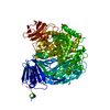 5jovC  5jowC  5joxC  5joyC  5jozC  5jp0C 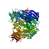 2xvgS S: Starting model for refinement C: citing same article ( |
|---|---|
| Similar structure data |


-Links
 PDBj
PDBj


- Assembly
| Deposited unit | 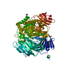
| ||||||||
|---|---|---|---|---|---|---|---|---|---|
| 1 |
| ||||||||
| Unit cell |
|
-Components
| #1: Protein | Mass: 109938.469 Da / Num. of mol.: 1 / Fragment: UNP residues 22-954 Source method: isolated from a genetically manipulated source Source: (gene. exp.) Bacteroides ovatus (bacteria) / Gene: BACOVA_02646 / Plasmid: pET-YSBL3C / Production host: Escherichia coli BL21(DE3) (bacteria) / Variant (production host): TUNER / References: UniProt: A7LXT0, alpha-D-xyloside xylohydrolase | ||
|---|---|---|---|
| #2: Chemical | ChemComp-NI / Nickel  Mass: 58.693 Da / Num. of mol.: 1 / Source method: obtained synthetically / Formula: Ni Mass: 58.693 Da / Num. of mol.: 1 / Source method: obtained synthetically / Formula: Ni | ||
| #3: Chemical | ChemComp-EDO / Ethylene glycol  Mass: 62.068 Da / Num. of mol.: 9 / Source method: obtained synthetically / Formula: C2H6O2 Mass: 62.068 Da / Num. of mol.: 9 / Source method: obtained synthetically / Formula: C2H6O2#4: Water | ChemComp-HOH / | Water Mass: 18.015 Da / Num. of mol.: 1644 / Source method: isolated from a natural source / Formula: H2O Mass: 18.015 Da / Num. of mol.: 1644 / Source method: isolated from a natural source / Formula: H2O |
-Experimental details
-Experiment
| Experiment | Method: X-RAY DIFFRACTION / Number of used crystals: 1 |
|---|
- Sample preparation
| Crystal | Density Matthews: 2.77 Å3/Da / Density % sol: 55.52 % |
|---|---|
| Crystal grow | Temperature: 292 K / Method: vapor diffusion, hanging drop / Details: 0.2 M potassium thiocyanate, 14-20% (w/v) PEG-3350 |
-Data collection
| Diffraction | Mean temperature: 100 K |
|---|---|
| Diffraction source | Source: SYNCHROTRON / Site: Diamond / Beamline: I04-1 / Wavelength: 0.92 Å |
| Detector | Type: DECTRIS PILATUS 2M / Detector: PIXEL / Date: Dec 12, 2013 |
| Radiation | Protocol: SINGLE WAVELENGTH / Monochromatic (M) / Laue (L): M / Scattering type: x-ray |
| Radiation wavelength | Wavelength: 0.92 Å / Relative weight: 1 |
| Reflection | Resolution: 1.5→48.35 Å / Num. obs: 187821 / % possible obs: 96.7 % / Redundancy: 7.5 % / CC1/2: 0.997 / Rmerge(I) obs: 0.124 / Net I/σ(I): 12.4 |
| Reflection shell | Resolution: 1.5→1.53 Å / Redundancy: 4.2 % / Rmerge(I) obs: 1.09 / Mean I/σ(I) obs: 1 / % possible all: 74.5 |
- Processing
| Software |
| ||||||||||||||||||||||||||||||||||||||||||||||||||||||||||||||||||||||||||||||||||||||||||||||||||||||||||||||||||||||||||||||||||||||||||||||||||||||||||||||||||||||||||||||||||||||
|---|---|---|---|---|---|---|---|---|---|---|---|---|---|---|---|---|---|---|---|---|---|---|---|---|---|---|---|---|---|---|---|---|---|---|---|---|---|---|---|---|---|---|---|---|---|---|---|---|---|---|---|---|---|---|---|---|---|---|---|---|---|---|---|---|---|---|---|---|---|---|---|---|---|---|---|---|---|---|---|---|---|---|---|---|---|---|---|---|---|---|---|---|---|---|---|---|---|---|---|---|---|---|---|---|---|---|---|---|---|---|---|---|---|---|---|---|---|---|---|---|---|---|---|---|---|---|---|---|---|---|---|---|---|---|---|---|---|---|---|---|---|---|---|---|---|---|---|---|---|---|---|---|---|---|---|---|---|---|---|---|---|---|---|---|---|---|---|---|---|---|---|---|---|---|---|---|---|---|---|---|---|---|---|
| Refinement | Method to determine structure: MOLECULAR REPLACEMENT Starting model: 2xvg Resolution: 1.5→48.35 Å / Cor.coef. Fo:Fc: 0.97 / Cor.coef. Fo:Fc free: 0.961 / SU B: 1.488 / SU ML: 0.051 / Cross valid method: THROUGHOUT / ESU R: 0.065 / ESU R Free: 0.066 / Details: HYDROGENS HAVE BEEN ADDED IN THE RIDING POSITIONS
| ||||||||||||||||||||||||||||||||||||||||||||||||||||||||||||||||||||||||||||||||||||||||||||||||||||||||||||||||||||||||||||||||||||||||||||||||||||||||||||||||||||||||||||||||||||||
| Solvent computation | Ion probe radii: 0.8 Å / Shrinkage radii: 0.8 Å / VDW probe radii: 1.2 Å | ||||||||||||||||||||||||||||||||||||||||||||||||||||||||||||||||||||||||||||||||||||||||||||||||||||||||||||||||||||||||||||||||||||||||||||||||||||||||||||||||||||||||||||||||||||||
| Displacement parameters | Biso mean: 14.903 Å2
| ||||||||||||||||||||||||||||||||||||||||||||||||||||||||||||||||||||||||||||||||||||||||||||||||||||||||||||||||||||||||||||||||||||||||||||||||||||||||||||||||||||||||||||||||||||||
| Refinement step | Cycle: 1 / Resolution: 1.5→48.35 Å
| ||||||||||||||||||||||||||||||||||||||||||||||||||||||||||||||||||||||||||||||||||||||||||||||||||||||||||||||||||||||||||||||||||||||||||||||||||||||||||||||||||||||||||||||||||||||
| Refine LS restraints |
|